ABG Interpretation: A Step-by-Step Guide for Medical Students
Arterial blood gas (ABG) interpretation is an essential clinical skill for medical students, residents, and healthcare professionals. An ABG can help identify acid-base disturbances, assess ventilation, evaluate oxygenation, and recognize potentially dangerous mixed disorders.
The key is to interpret the results systematically rather than trying to memorize every possible combination of values. This guide explains a practical approach to ABG interpretation, including normal ranges, compensation rules, anion gap assessment, worked examples, and practice cases.
1. Understanding ABG Values
An arterial blood gas measures several aspects of respiratory and metabolic function. The most important values for initial interpretation are pH, partial pressure of carbon dioxide (PaCO₂), and bicarbonate (HCO₃⁻).
pH: Is the blood acidemic or alkalemic?
Blood pH reflects the balance between acids and bases. A low pH indicates acidemia, while a high pH indicates alkalemia. These terms describe the measured blood state; they are not necessarily the same as the name of the underlying primary disorder.
PaCO₂: The respiratory component
Carbon dioxide behaves as an acid through its relationship with carbonic acid. Increased PaCO₂ tends to lower pH, while decreased PaCO₂ tends to raise pH. The lungs regulate PaCO₂ by changing alveolar ventilation.
HCO₃⁻: The metabolic component
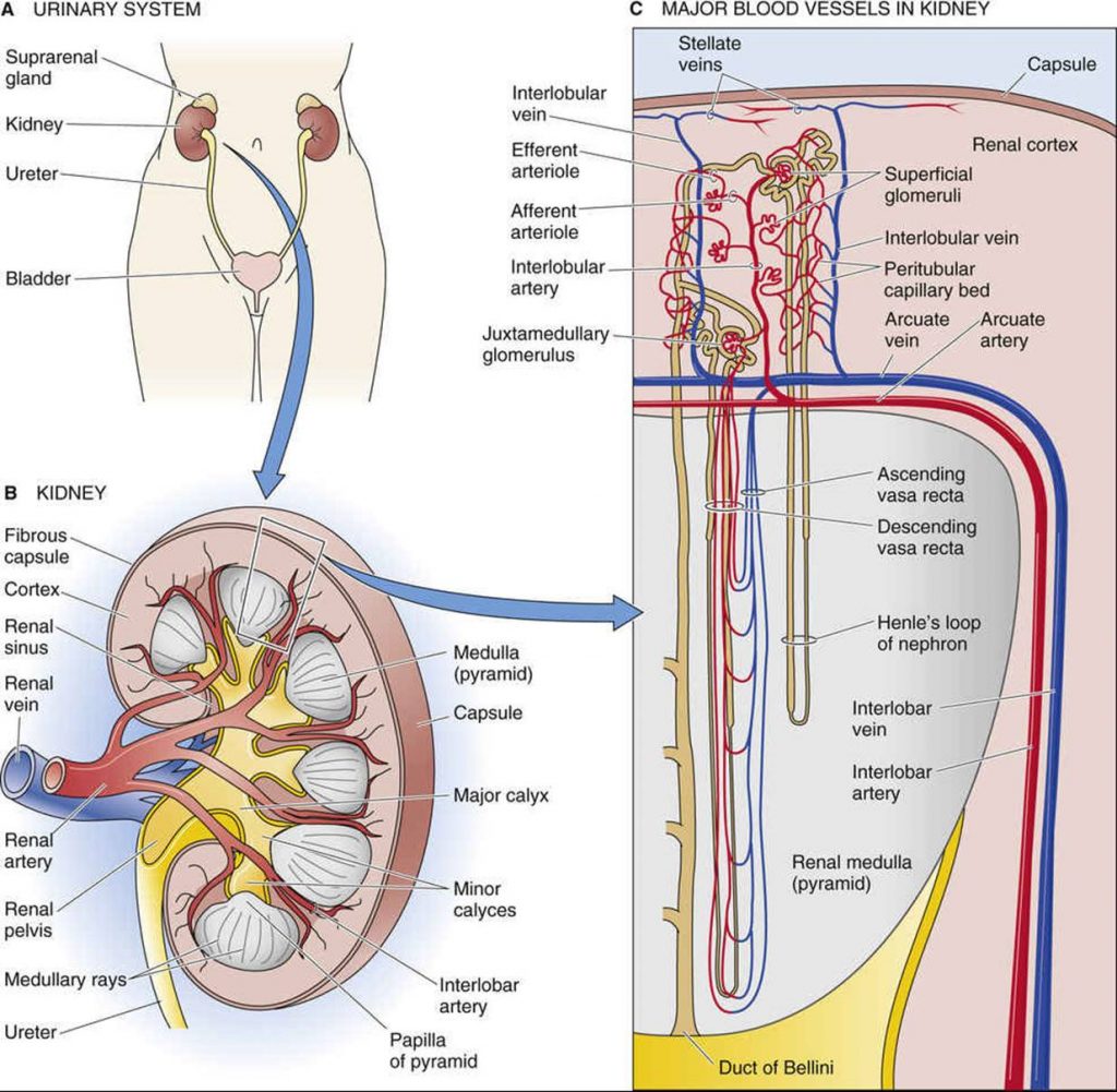
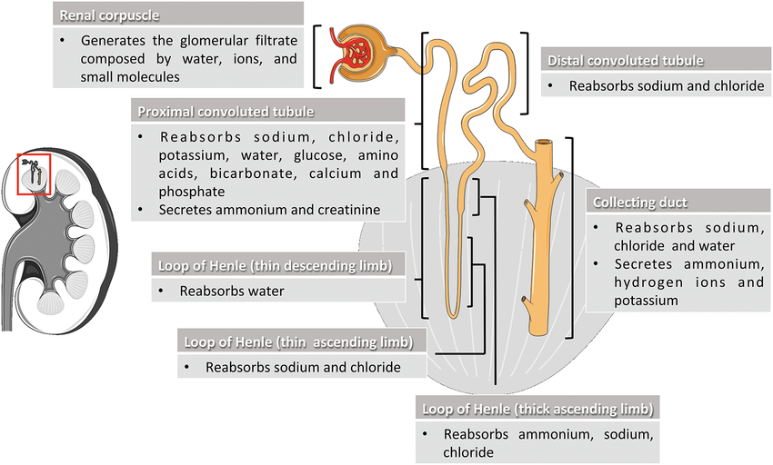
Bicarbonate is the principal measured component used to assess the metabolic side of the acid-base system. A decrease in bicarbonate tends to lower pH, while an increase tends to raise pH. The kidneys play a major role in regulating bicarbonate and acid excretion.
PaO₂ and oxygen saturation
PaO₂ reflects the partial pressure of dissolved oxygen in arterial blood. It helps assess oxygenation, but it does not directly represent the total oxygen content of the blood. Hemoglobin concentration and oxygen saturation are also important when evaluating oxygen delivery.
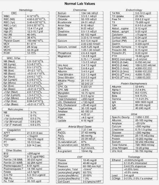
2. Normal ABG Values
The following are commonly used reference ranges for adults at sea level. Actual ranges depend on the laboratory, clinical setting, age, and circumstances of sampling.
| Parameter | Common reference range | Clinical meaning |
|---|---|---|
| pH | 7.35–7.45 | Overall acid-base status |
| PaCO₂ | 35–45 mmHg | Respiratory component |
| HCO₃⁻ | 22–26 mEq/L | Metabolic component |
| PaO₂ | Approximately 80–100 mmHg | Arterial oxygenation; varies with age and altitude |
| Oxygen saturation | Often 95–100% | Hemoglobin oxygen saturation; interpret in context |
These values are general teaching references, not universal cutoffs. Always check the reporting laboratory's reference intervals and consider the patient's baseline.
3. The Five-Step ABG Interpretation Method
Use the same sequence for every ABG. A structured approach makes it easier to identify the primary disorder and recognize when more than one process is present.
-
Determine the pH.
A pH below 7.35 indicates acidemia. A pH above 7.45 indicates alkalemia. A pH within the reference range does not exclude a significant acid-base disorder because opposing processes can coexist.
-
Identify the primary respiratory or metabolic process.
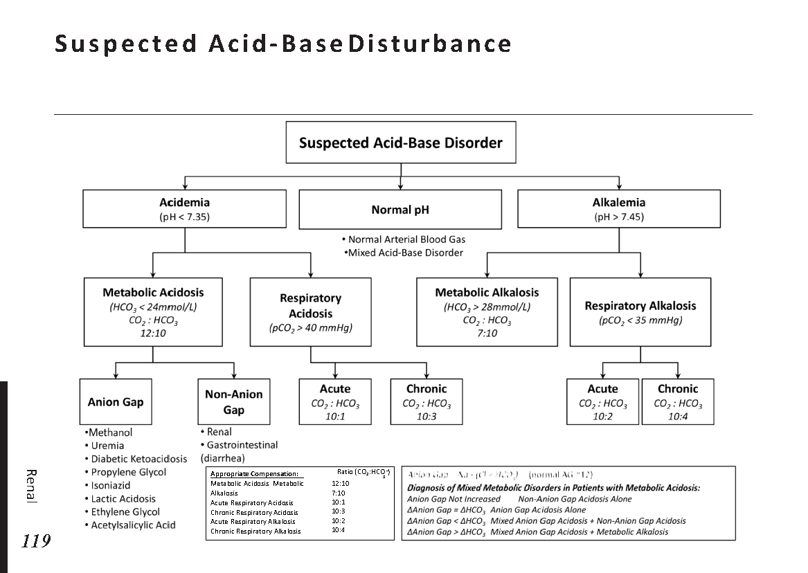
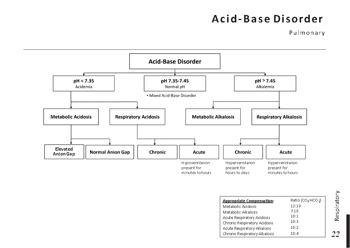
In acidemia, a high PaCO₂ suggests a respiratory contribution, while a low HCO₃⁻ suggests a metabolic contribution. In alkalemia, a low PaCO₂ suggests a respiratory contribution, while a high HCO₃⁻ suggests a metabolic contribution.
-
Check whether compensation is appropriate.
Use the expected compensation rules for the suspected primary disorder. A response outside the expected range raises concern for an additional acid-base process.
-
Calculate the anion gap when metabolic acidosis is present.
The anion gap helps distinguish high-anion-gap metabolic acidosis from normal-anion-gap metabolic acidosis. Albumin concentration can significantly affect interpretation.
-
Evaluate oxygenation and the clinical context.
Review PaO₂, oxygen saturation, inspired oxygen concentration, respiratory support, vital signs, symptoms, and relevant laboratory findings. An ABG must be interpreted alongside the patient, not in isolation.
A quick example
Suppose the ABG shows pH 7.25, PaCO₂ 60 mmHg, and HCO₃⁻ 26 mEq/L.
- The pH indicates acidemia.
- The PaCO₂ is elevated and explains the direction of the pH.
- The primary disorder is respiratory acidosis.
- Assess whether the bicarbonate response is appropriate for an acute or chronic process.
4. The Four Primary Acid-Base Disorders
Respiratory acidosis
Respiratory acidosis occurs when inadequate alveolar ventilation causes carbon dioxide retention. The increased PaCO₂ lowers blood pH.
Common causes include:
- Opioid or sedative-related respiratory depression.
- Severe obstructive lung disease with ventilatory failure.
- Neuromuscular weakness affecting ventilation.
- Severe airway obstruction or respiratory fatigue.
- Chest wall or central nervous system disorders that impair ventilation.
The kidneys compensate by retaining bicarbonate and increasing net acid excretion. This response takes time, so chronic respiratory acidosis generally produces a larger bicarbonate increase than acute respiratory acidosis.
Respiratory alkalosis
Respiratory alkalosis results from excessive alveolar ventilation and a reduction in PaCO₂, which raises pH.
Common causes include:
- Hypoxemia and pulmonary disease.
- Pulmonary embolism.
- Pain, anxiety, or acute stress.
- Sepsis and other systemic illnesses.
- Pregnancy and certain central nervous system conditions.
- Excessive mechanical ventilation.
The kidneys compensate by decreasing bicarbonate reabsorption and increasing bicarbonate excretion. The degree of compensation depends on the duration of the respiratory disturbance.
Metabolic acidosis
Metabolic acidosis is characterized by a primary reduction in bicarbonate, which lowers pH. Respiratory compensation generally occurs through increased ventilation and a reduction in PaCO₂.
Important causes include:
- Lactic acidosis associated with shock, hypoperfusion, or other conditions.
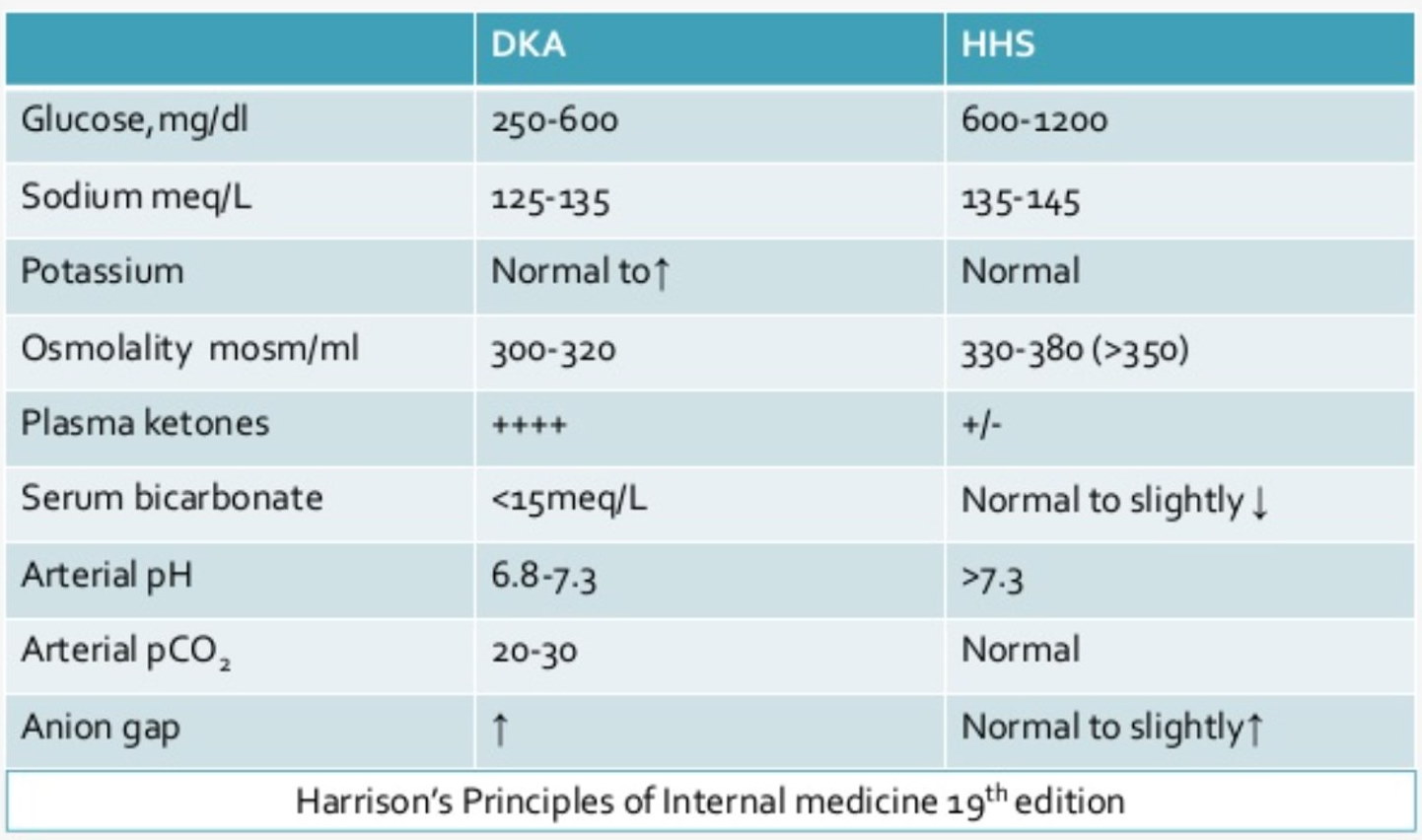
- Diabetic ketoacidosis and other forms of ketoacidosis.
- Renal impairment with reduced acid excretion.
- Diarrhea or other causes of bicarbonate loss.
- Renal tubular acidosis.
- Selected medication or toxic exposures.
Once metabolic acidosis is identified, calculate the anion gap and investigate the cause. The clinical assessment may require serum electrolytes, lactate, ketones, renal function, glucose, and targeted toxicology testing.
Metabolic alkalosis
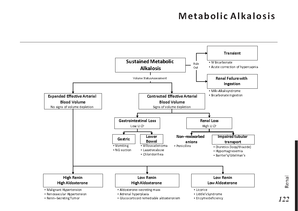
Metabolic alkalosis is characterized by a primary increase in bicarbonate and an increase in pH. The respiratory system compensates through reduced ventilation, which increases PaCO₂. This response is limited by the need to maintain adequate oxygenation.
Common causes include:
- Vomiting or nasogastric suction causing gastric acid loss.
- Diuretic use.
- Mineralocorticoid excess.
- Hypokalemia and chloride depletion.
- Alkali administration in susceptible patients.
Assess volume status, medications, potassium, chloride, renal function, and urine chloride when clinically appropriate. Management depends on the underlying cause.
5. Expected Compensation in ABG Interpretation
Compensation is the body's response to a primary acid-base disturbance. The lungs respond rapidly to metabolic disturbances, whereas renal compensation for respiratory disorders develops over hours to days.
Compensation does not usually normalize the pH completely. If measured values differ substantially from expected compensation, consider a mixed acid-base disorder.
Winter's formula for metabolic acidosis
This formula estimates the expected respiratory response in metabolic acidosis.
- If the measured PaCO₂ is within the expected range, respiratory compensation is broadly appropriate.
- If the measured PaCO₂ is higher than expected, a concurrent respiratory acidosis may be present.
- If the measured PaCO₂ is lower than expected, a concurrent respiratory alkalosis may be present.
Respiratory acidosis: acute versus chronic
For each 10 mmHg increase in PaCO₂ above 40 mmHg, bicarbonate is expected to increase approximately:
| Process | Expected HCO₃⁻ change |
|---|---|
| Acute respiratory acidosis | Increase by about 1 mEq/L |
| Chronic respiratory acidosis | Increase by about 3.5–4 mEq/L |
Respiratory alkalosis: acute versus chronic
For each 10 mmHg decrease in PaCO₂ below 40 mmHg, bicarbonate is expected to decrease approximately:
| Process | Expected HCO₃⁻ change |
|---|---|
| Acute respiratory alkalosis | Decrease by about 2 mEq/L |
| Chronic respiratory alkalosis | Decrease by about 4–5 mEq/L |
Metabolic alkalosis
In metabolic alkalosis, PaCO₂ usually increases by approximately 0.5–0.7 mmHg for each 1 mEq/L increase in bicarbonate above 24 mEq/L. Estimates vary, and the response may be limited by hypoxemia or other respiratory constraints.
6. Anion Gap and Mixed Acid-Base Disorders
How to calculate the anion gap
The anion gap is commonly used to classify metabolic acidosis. When potassium is excluded, the standard formula is:
A typical reference range is approximately 8–12 mEq/L when potassium is excluded, but the laboratory's own reference range should be used. Different measurement methods can produce different ranges.
High-anion-gap metabolic acidosis
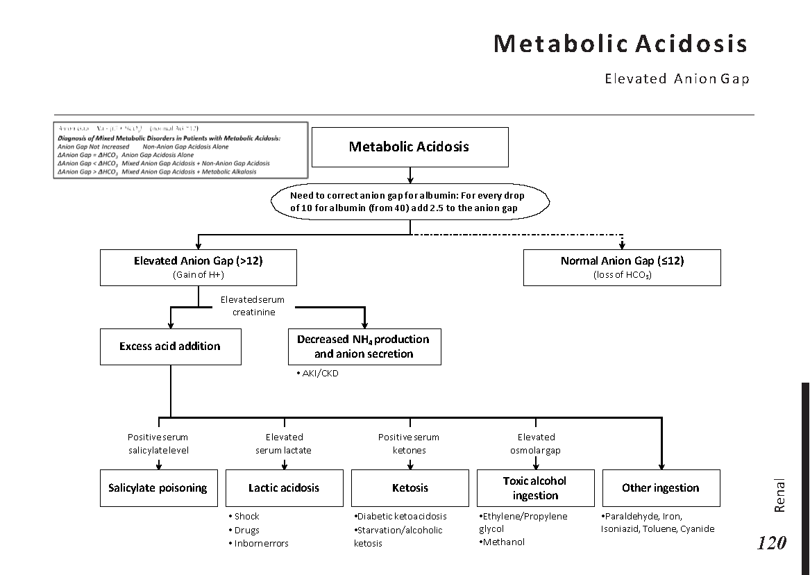
A high anion gap suggests the accumulation of unmeasured anions. Important causes include:
- Lactic acidosis.
- Ketoacidosis, including diabetic, alcoholic, and starvation-related forms.
- Advanced renal failure.
- Selected toxic alcohols and medication-related poisonings.
- Other causes of accumulation of organic acids.
The cause cannot be determined from the anion gap alone. Use the patient's history, examination, medication exposure, renal function, lactate, ketones, and other targeted tests.
Correcting the anion gap for albumin
Albumin is a major unmeasured anion. Low albumin can make a clinically important high anion gap appear less pronounced.
This is a commonly used approximation. Interpret the result in light of the laboratory method and the patient's clinical context.
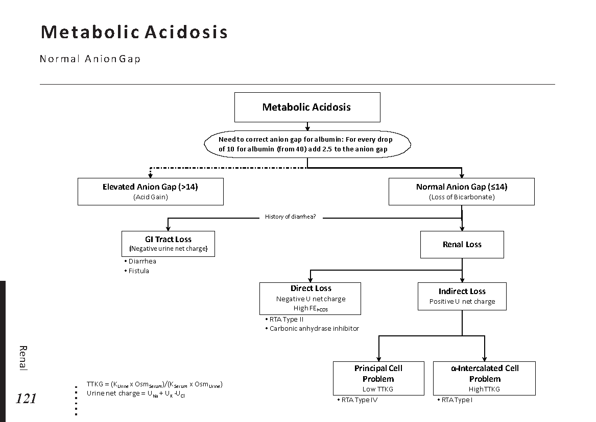
Normal-anion-gap metabolic acidosis
In normal-anion-gap metabolic acidosis, bicarbonate decreases while chloride commonly increases. This pattern is sometimes called hyperchloremic metabolic acidosis.
Potential causes include:
- Gastrointestinal bicarbonate loss, particularly diarrhea.
- Renal tubular acidosis.
- Selected medications.
- Large volumes of chloride-rich intravenous fluid in some settings.
Recognizing mixed acid-base disorders
A mixed disorder occurs when two or more primary acid-base processes coexist. It should be suspected when compensation is outside the expected range or when the clinical picture does not fit the measured values.
Example: metabolic acidosis with respiratory acidosis
Consider pH 7.10, HCO₃⁻ 12 mEq/L, and PaCO₂ 40 mmHg.
Winter's formula predicts an expected PaCO₂ of:
The measured PaCO₂ of 40 mmHg is substantially higher than expected. This suggests a concurrent respiratory acidosis in addition to the metabolic acidosis.
Delta ratio
In high-anion-gap metabolic acidosis, the delta ratio can help identify an additional metabolic process. One commonly used formula is:
Use a laboratory-appropriate normal anion gap, commonly around 12 mEq/L when potassium is excluded. Interpretation is approximate: a relatively low ratio may suggest an additional normal-anion-gap metabolic acidosis, while a relatively high ratio may suggest a concurrent metabolic alkalosis or another process. Cutoffs vary, and the delta ratio should never replace full clinical interpretation.
7. Assessing Oxygenation on an ABG
Acid-base interpretation is only part of ABG analysis. Oxygenation should be assessed separately, taking into account the patient's oxygen delivery settings, altitude, age, and clinical condition.
PaO₂
PaO₂ is the partial pressure of oxygen dissolved in arterial blood. A low value can indicate impaired oxygenation, but interpretation depends on the inspired oxygen concentration and the clinical setting.
PaO₂/FiO₂ ratio
For example, if PaO₂ is 80 mmHg while the patient receives an FiO₂ of 0.40, the P/F ratio is 200.
The ratio is used to assess the severity of impaired oxygenation in appropriate clinical contexts. Its interpretation depends on factors including ventilatory support and the diagnostic criteria being used. It should not be used alone to diagnose acute respiratory distress syndrome (ARDS).
Alveolar-arterial oxygen gradient
The A–a gradient compares estimated alveolar oxygen pressure with measured arterial oxygen pressure. It can help distinguish different mechanisms of hypoxemia when interpreted in context.
At sea level, the alveolar gas equation is commonly approximated as:
This approximation assumes sea-level atmospheric pressure, water vapor pressure of 47 mmHg, and a respiratory quotient of 0.8. Adjust the equation for different environmental conditions when necessary.
A normal A–a gradient generally points toward causes such as hypoventilation or low inspired oxygen, whereas an increased gradient may occur with ventilation-perfusion mismatch, diffusion impairment, or right-to-left shunting. Expected values vary with age and inspired oxygen concentration.
8. Worked Clinical Cases
Try interpreting each case before opening the explanation. Focus on pH first, then identify the primary process and assess compensation.
Case 1: Vomiting and weakness
A patient presents with several days of vomiting. The ABG shows:
- pH: 7.50
- PaCO₂: 48 mmHg
- HCO₃⁻: 36 mEq/L
Reveal the interpretation
Primary disorder: Metabolic alkalosis.
The pH is elevated and bicarbonate is increased. PaCO₂ is also increased, consistent with respiratory compensation. Vomiting can cause loss of gastric hydrochloric acid and contribute to metabolic alkalosis.
Further assessment may include volume status, potassium, chloride, renal function, and the cause of the vomiting.
Case 2: Severe hyperglycemia
A patient with diabetes presents with nausea, dehydration, and deep breathing. The ABG shows:
- pH: 7.20
- PaCO₂: 24 mmHg
- HCO₃⁻: 9 mEq/L
Reveal the interpretation
Primary disorder: Metabolic acidosis.
The low pH and low bicarbonate indicate metabolic acidosis. Winter's formula predicts a PaCO₂ of approximately 21.5 ± 2 mmHg. The measured PaCO₂ of 24 mmHg is at the upper edge of the expected range, supporting broadly appropriate respiratory compensation.
Given the clinical history, diabetic ketoacidosis is an important possibility, but it requires assessment of glucose, ketones, electrolytes, and other diagnostic criteria.
Case 3: Reduced consciousness after sedative use
A patient is drowsy with slow breathing. The ABG shows:
- pH: 7.24
- PaCO₂: 60 mmHg
- HCO₃⁻: 25 mEq/L
Reveal the interpretation
Likely primary disorder: Acute respiratory acidosis.
The pH is low and PaCO₂ is elevated. Bicarbonate is close to normal, which is compatible with a relatively acute respiratory disturbance. Sedative-related hypoventilation is a possible cause.
A real patient with reduced consciousness and suspected hypoventilation requires urgent assessment of airway protection, breathing, oxygenation, and the underlying cause.
Case 4: Tachypnea with sepsis
A patient with suspected sepsis is breathing rapidly. The ABG shows:
- pH: 7.48
- PaCO₂: 28 mmHg
- HCO₃⁻: 20 mEq/L
Reveal the interpretation
Likely primary disorder: Respiratory alkalosis.
The elevated pH and reduced PaCO₂ support respiratory alkalosis. The bicarbonate reduction may reflect compensation, but the duration of the process and the full clinical context matter.
Sepsis may cause respiratory alkalosis and may also cause lactic metabolic acidosis. Assess lactate, electrolytes, anion gap, perfusion, and other clinical findings before concluding that only one process is present.
9. Test Your ABG Interpretation Skills
Choose the best answer for each question, then select Check answers to review your results.
10. Common ABG Interpretation Mistakes
1. Looking at PaCO₂ before checking the pH
Start with the pH to establish whether acidemia or alkalemia is present. Then evaluate PaCO₂ and bicarbonate to identify the primary process.
2. Assuming a normal pH means there is no disorder
A normal pH can occur when two primary processes oppose one another. Check whether the PaCO₂ and bicarbonate are abnormal and whether compensation is appropriate.
3. Ignoring expected compensation
A change in the second variable is not automatically compensation. Compare the measured response with the expected response to identify possible mixed disorders.
4. Forgetting the anion gap
In metabolic acidosis, the anion gap provides important diagnostic information. A low albumin concentration can conceal an elevated gap.
5. Confusing oxygenation with ventilation
PaCO₂ primarily informs the assessment of ventilation, whereas PaO₂ informs the assessment of oxygenation. A patient can have a problem with one, the other, or both.
6. Interpreting numbers without the clinical history
The same ABG pattern may have different causes depending on the patient's medications, medical history, vital signs, laboratory findings, oxygen support, and duration of illness.
11. References and Further Reading
Use established clinical references to deepen your understanding of acid-base physiology, compensation, and arterial blood gas analysis. The following resources provide useful starting points:
- Merck Manual Professional Edition — clinical reference material on acid-base disorders and their evaluation.
- NCBI Bookshelf — biomedical books and clinical reference chapters.
- PubMed — peer-reviewed research and reviews on acid-base physiology and ABG interpretation.
Reference ranges and compensation estimates in this article are educational approximations. Verify clinical decisions against current guidelines, laboratory standards, and appropriate specialist advice.
Practice with the Medicogenic ABG Calculator
After working through the interpretation steps, use our existing ABG Calculator to support your practice. Calculate relevant values when available, then interpret the results independently using the method explained in this guide.
 Menstrual cycle physiology: