Acute Pancreatitis: Diagnosis, Severity, and Initial Management
Acute pancreatitis is an inflammatory condition of the pancreas that can range from a mild, self-limited illness to a life-threatening disease involving respiratory failure, circulatory collapse, or infected pancreatic necrosis. Recognizing the diagnosis is only the first step: clinicians must also identify the underlying cause, assess severity, and provide appropriate early treatment.
For medical students and junior clinicians, the most useful approach is to connect the patient's symptoms and laboratory findings with the decisions that change management. This guide explains that approach through practical clinical reasoning rather than a list of isolated facts.
1. What happens in acute pancreatitis?
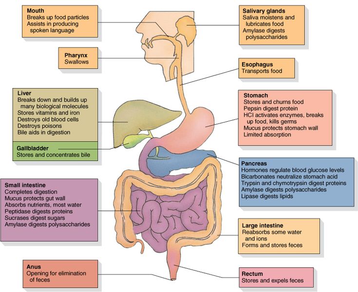
The pancreas produces digestive enzymes and hormones, including insulin and glucagon. In acute pancreatitis, premature activation of digestive enzymes within the pancreas contributes to tissue injury and an inflammatory response.
In mild disease, inflammation remains relatively limited and resolves with supportive care. In more serious cases, the inflammatory response can affect the entire body, impair circulation, and damage distant organs. Pancreatic tissue may also develop areas of necrosis.
The severity of the illness therefore depends not simply on pancreatic inflammation itself, but also on whether the patient develops organ dysfunction and local complications.
2. How is acute pancreatitis diagnosed?
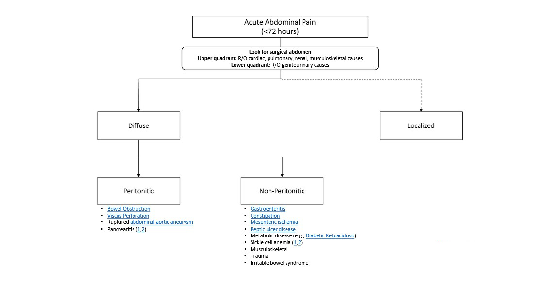
The diagnosis is generally established when at least two of the following three criteria are present:
- Characteristic abdominal pain, typically persistent epigastric pain that may radiate to the back.
- Serum lipase or amylase at least three times the upper limit of normal.
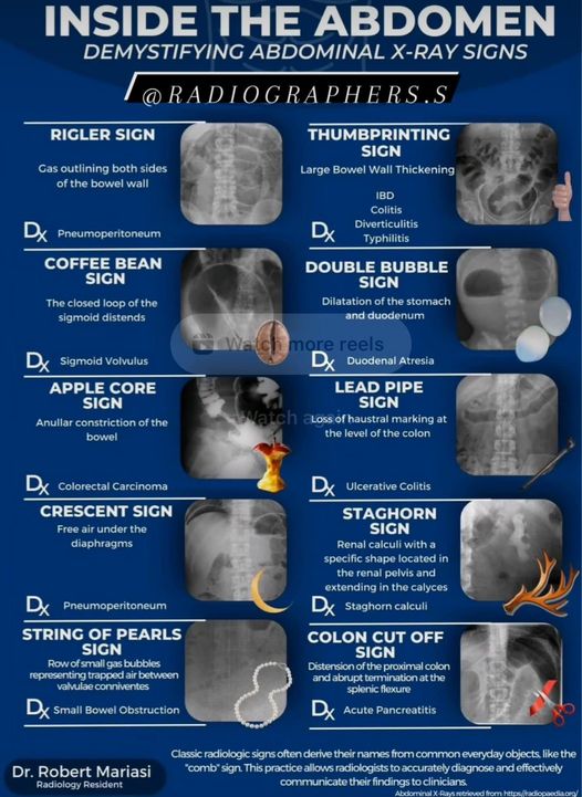
- Characteristic findings of acute pancreatitis on abdominal imaging.
Not every patient needs imaging to confirm the diagnosis. If the pain is characteristic and lipase is sufficiently elevated, the diagnosis can usually be made without immediate CT imaging.
Why is lipase usually preferred?
Serum lipase is generally preferred to amylase because it is more specific for pancreatic injury and tends to remain elevated longer. However, an elevated lipase alone does not prove acute pancreatitis. Other conditions, including renal impairment and some intra-abdominal illnesses, can also increase lipase.
3. Recognizing the typical presentation
Common clinical features include:
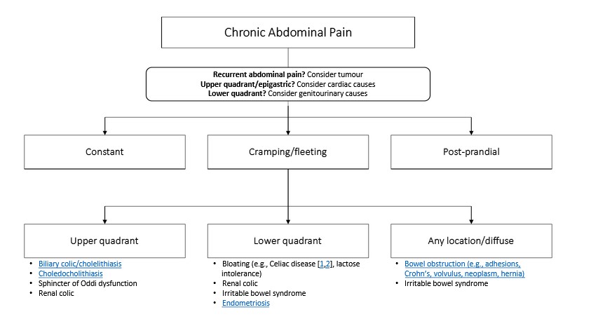
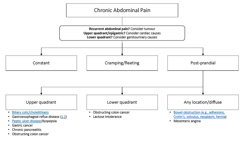
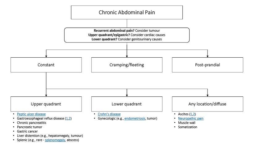
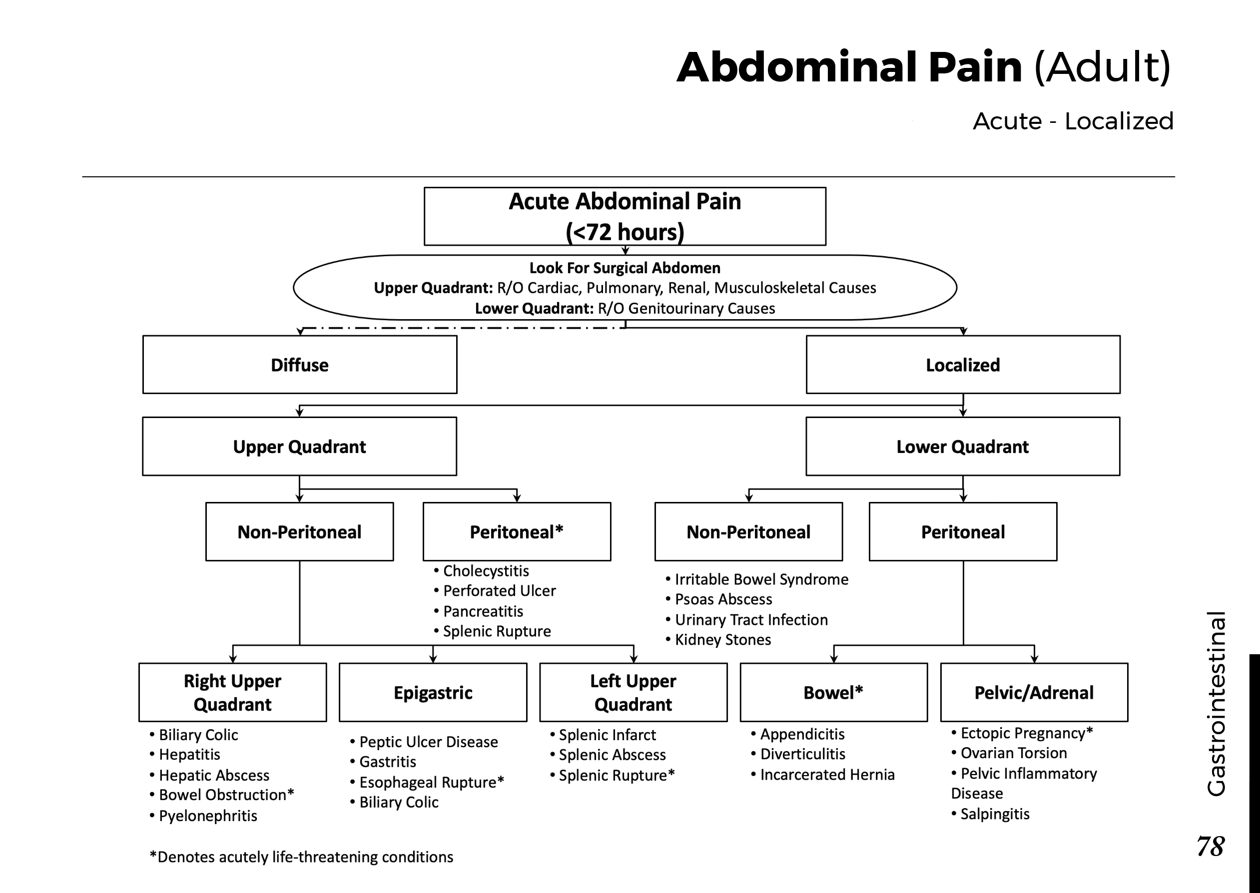
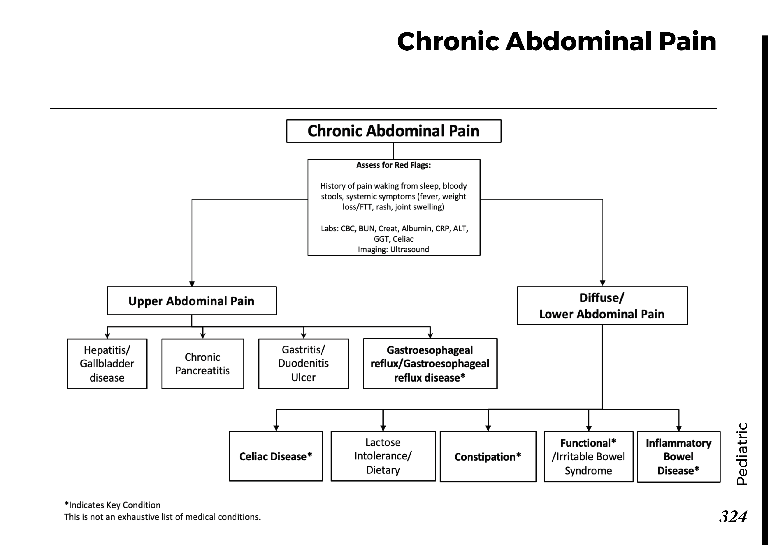
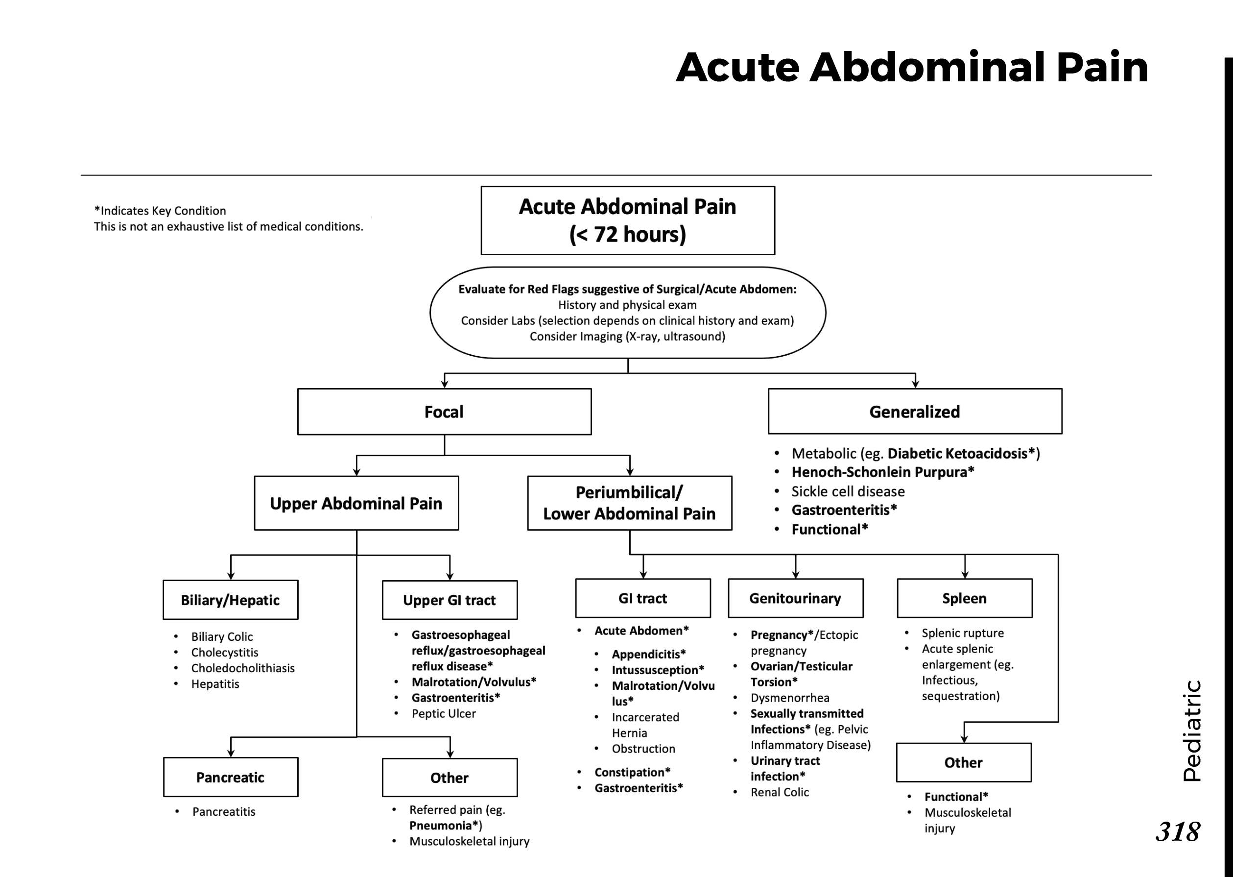
- Persistent upper abdominal, usually epigastric, pain.
- Pain that may radiate to the back.
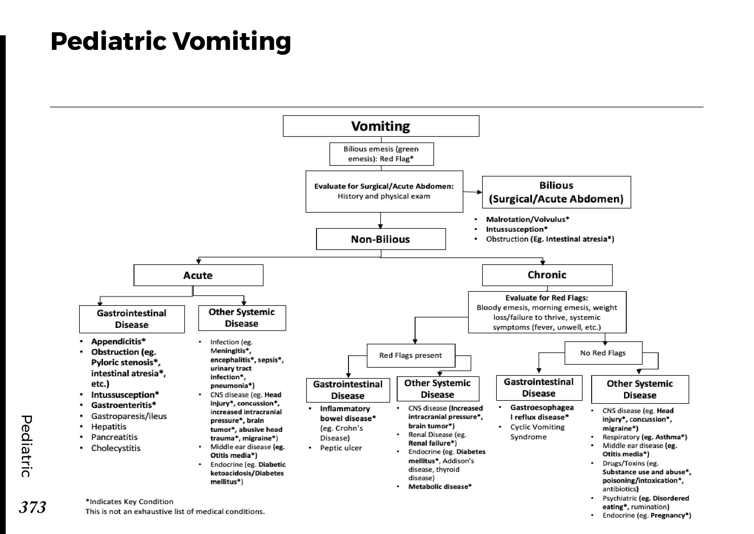
- Nausea and vomiting.
- Abdominal tenderness and reduced tolerance of oral intake.
- Tachycardia, fever, or dehydration in more significant illness.
Severe cases may present with hypotension, hypoxemia, oliguria, altered mental status, or other evidence of organ dysfunction. These findings require urgent assessment and treatment.
A physical examination can be relatively nonspecific. The absence of dramatic abdominal findings does not exclude pancreatitis, while marked guarding or rigidity should prompt evaluation for other serious abdominal pathology as well.
4. The major causes of acute pancreatitis
Identifying the cause matters because some patients need a specific intervention to prevent recurrence or treat an underlying obstruction.
| Cause | Clinical clues | Important next step |
|---|---|---|
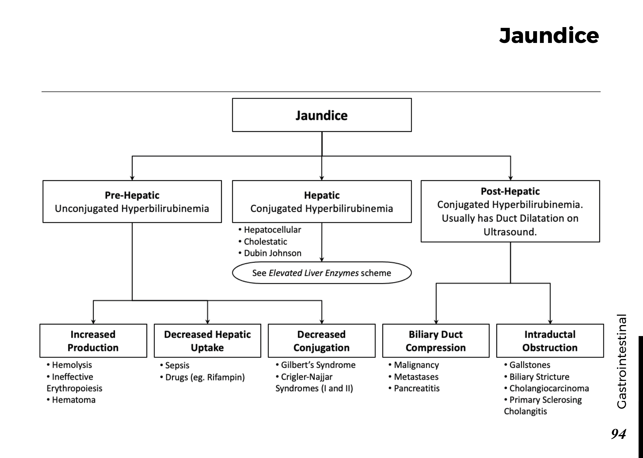
| Gallstones | Right upper quadrant symptoms, gallstones on ultrasound, or abnormal liver tests. | Perform right upper quadrant ultrasound and assess for biliary obstruction. |
| Alcohol | Relevant alcohol exposure or a compatible previous history. | Take a nonjudgmental history and assess other contributing causes. |
| Hypertriglyceridemia | Markedly elevated triglycerides or a history of severe lipid abnormalities. | Measure triglycerides early, particularly if the cause is unclear. |
| Medications | Recent exposure to a drug associated with pancreatitis. | Review prescribed medicines and recent changes; consider alternative causes before attributing causality. |
| Other causes | Recent procedures, hypercalcemia, trauma, genetic factors, or no identified cause. | Investigate according to the clinical history and initial findings. |
Gallstones and alcohol are common causes. If neither is evident, further evaluation should be guided by the patient's age, history, laboratory results, and imaging findings. A first episode without a clear cause should not automatically be labeled idiopathic without appropriate assessment.
5. Initial investigations: what does each test tell you?
Blood tests
- Lipase: Helps establish the diagnosis in the appropriate clinical context.
- Complete blood count: May show leukocytosis or hemoconcentration.
- Urea, creatinine, and electrolytes: Help assess hydration, renal function, and metabolic disturbances.
- Liver tests and bilirubin: May suggest a biliary cause or persistent bile duct obstruction.
- Glucose: May be elevated because of stress and pancreatic dysfunction.
- Calcium and triglycerides: Help investigate potential causes when indicated.
No single blood test reliably predicts the entire clinical course. Trends in renal function, urea, hematocrit, vital signs, and oxygenation can be more useful when interpreted together and reassessed over time.
Imaging
Right upper quadrant ultrasound is useful for detecting gallstones and assessing the biliary system. It is commonly part of the initial evaluation when gallstone pancreatitis is possible.
Contrast-enhanced CT is not routinely required immediately when the diagnosis is already clear and the patient is improving. It becomes useful when the diagnosis remains uncertain, an alternative condition is suspected, or the patient fails to improve or deteriorates. Imaging to evaluate necrosis is often more informative after the early phase of illness, depending on the clinical question.
MRI or MRCP and endoscopic ultrasound may be useful in selected patients when common bile duct stones or another pancreaticobiliary cause remains suspected.
6. Assessing severity: the distinction that changes management
Acute pancreatitis is classified according to the revised Atlanta framework:
| Category | Definition | Clinical meaning |
|---|---|---|
| Mild | No organ failure and no local or systemic complications. | Often resolves with supportive treatment and has a relatively short hospital course. |
| Moderately severe | Transient organ failure lasting no more than 48 hours and/or local or systemic complications without persistent organ failure. | Requires closer monitoring according to the patient's condition. |
| Severe | Persistent organ failure lasting more than 48 hours. | Associated with a substantially higher risk of complications and death. |
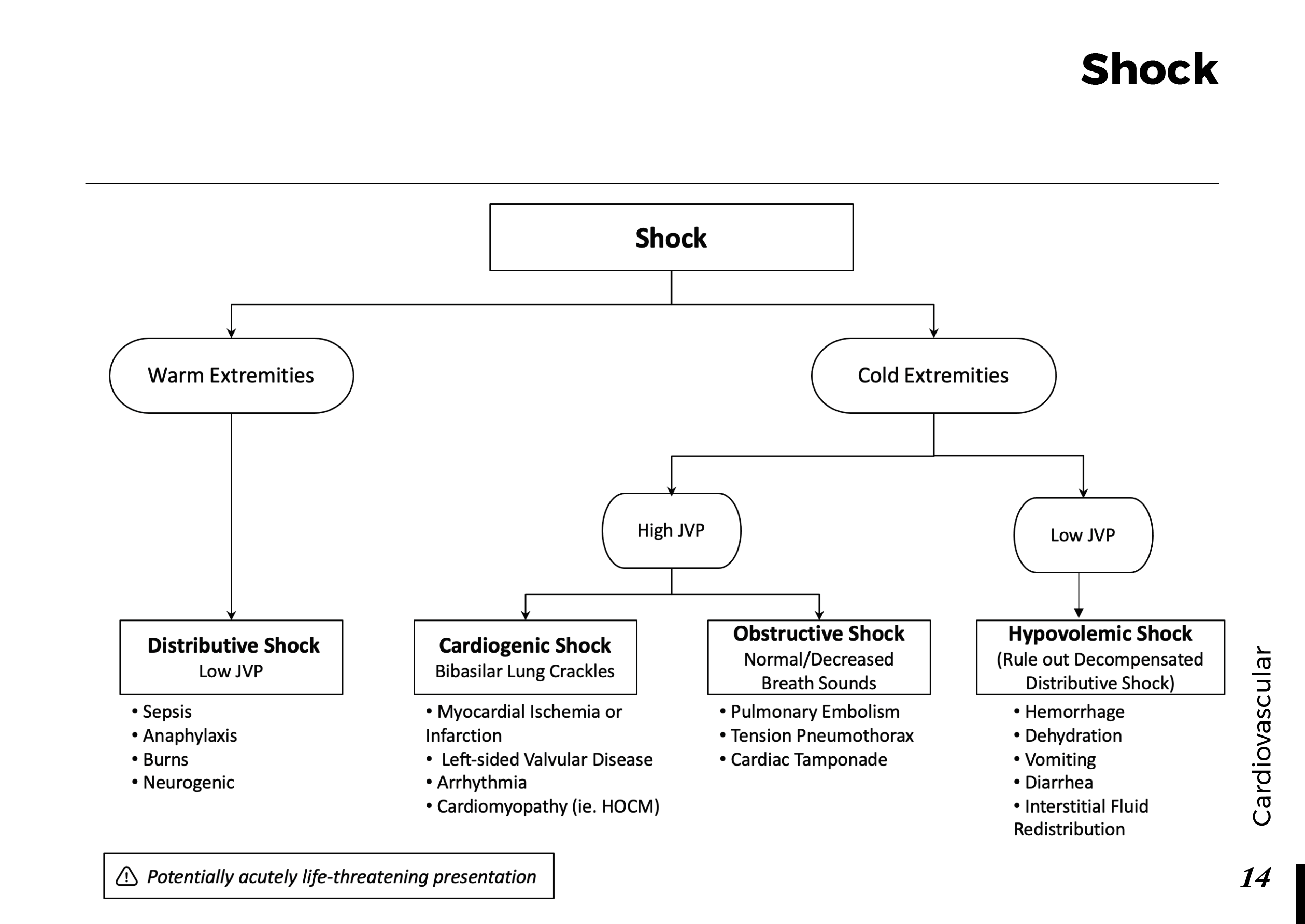
Organ failure may involve the respiratory, cardiovascular, or renal systems. Clinical deterioration, persistent hypotension, worsening oxygenation, or declining urine output should prompt urgent reassessment and escalation of care.
Scores such as BISAP and APACHE II can support risk assessment, but they do not replace clinical judgment. Severity can evolve during the first several days, so a reassuring initial assessment does not eliminate the need for reassessment.
7. Initial management: what should happen first?
Step 1: Assess and stabilize
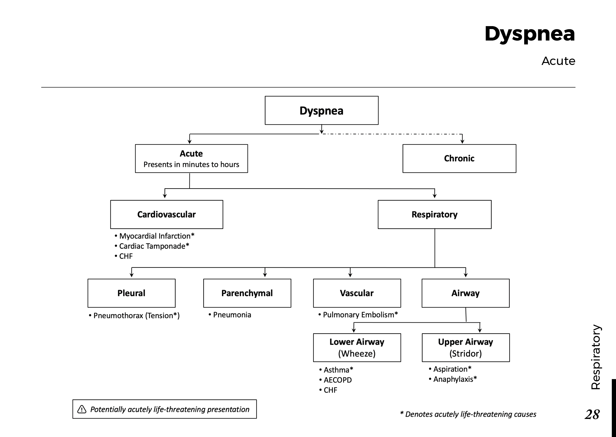
Begin with airway, breathing, circulation, vital signs, oxygenation, mental status, and urine output. Identify shock, respiratory compromise, or other organ dysfunction promptly. Patients with significant instability may need a higher level of monitoring or intensive care.
Step 2: Give appropriately monitored fluids
Intravenous crystalloid fluids are used when needed to correct hypovolemia and support perfusion. Lactated Ringer's solution is commonly preferred when appropriate. Fluid administration should be individualized, with frequent reassessment of blood pressure, heart rate, urine output, renal function, and signs of fluid overload.
Excessive fluid administration can be harmful, particularly in patients with cardiac or renal dysfunction. A fixed, aggressive fluid strategy should not be applied indiscriminately to every patient.
Step 3: Treat pain and nausea
Provide appropriate analgesia and antiemetics according to the patient's condition and local protocols. Adequate pain control is an important part of supportive management, not an optional extra.
Step 4: Resume nutrition early when tolerated
Patients with mild acute pancreatitis should generally resume oral feeding as tolerated rather than being kept fasting until pancreatic enzymes normalize. A low-fat solid diet may be suitable initially. If oral feeding is not possible, enteral nutrition is generally preferred over parenteral nutrition when nutritional support is needed.
Step 5: Avoid routine prophylactic antibiotics
Antibiotics are not routinely indicated for uncomplicated acute pancreatitis or sterile pancreatic necrosis. They should be used when there is a suspected or confirmed infection or another clear indication. Infected necrosis requires specialist assessment and an individualized treatment plan.
8. When is a procedure necessary?
Most patients do not need an immediate pancreatic procedure. Interventions depend on the underlying cause and the presence of complications.
- Gallstone pancreatitis with cholangitis: Urgent biliary evaluation and ERCP are generally indicated.
- Persistent common bile duct obstruction: Further assessment and possible therapeutic ERCP may be required.
- Mild gallstone pancreatitis: Cholecystectomy during the same admission is generally recommended when feasible to reduce recurrence.
- Pancreatic necrosis or collections: Management depends on symptoms, infection, maturity of the collection, and clinical stability. Many interventions can be delayed when clinically safe.
ERCP is not routinely needed for every patient with gallstone pancreatitis if there is no cholangitis or evidence of ongoing biliary obstruction.
9. Clinical cases: apply the reasoning
Case 1: The classic presentation
A 42-year-old patient presents with persistent epigastric pain radiating to the back and repeated vomiting. Serum lipase is eight times the upper limit of normal. The patient is hemodynamically stable.
Question: Is CT required immediately to confirm the diagnosis?
Case 2: The patient who may be deteriorating
A patient admitted with acute pancreatitis develops increasing oxygen requirements, persistent hypotension, and declining urine output.
Question: What should concern you most?
Case 3: A biliary emergency
A patient has acute pancreatitis, fever, jaundice, right upper quadrant discomfort, and laboratory findings suggesting biliary obstruction.
Question: Which complication must be considered, and what intervention may be needed?
Case 4: Feeding after improvement
A patient with mild acute pancreatitis has improving pain, no persistent vomiting, and can tolerate oral intake. The lipase remains elevated.
Question: Must oral feeding wait until lipase returns to normal?
10. Common mistakes to avoid
- Ordering immediate CT in every patient, even when the diagnosis is clear and the patient is improving.
- Using the height of the lipase elevation to determine disease severity.
- Giving excessive intravenous fluids without reassessment.
- Keeping every patient fasting until pancreatic enzymes normalize.
- Prescribing prophylactic antibiotics for sterile pancreatitis.
- Performing ERCP routinely in all cases of gallstone pancreatitis.
- Failing to investigate the cause and plan prevention of recurrence.
11. Frequently asked questions
Can acute pancreatitis occur with a normal lipase?
Yes, although this is uncommon. The timing of testing, certain underlying conditions, and the clinical context may affect enzyme levels. If suspicion remains high, reassess the diagnostic criteria and consider appropriate imaging or alternative diagnoses.
Does a higher lipase mean more severe pancreatitis?
No. Lipase is primarily a diagnostic marker. Clinical severity is assessed using organ function, complications, vital signs, laboratory trends, and the patient's course over time.
Are antibiotics routinely needed?
No. They are not recommended routinely for uncomplicated acute pancreatitis or sterile necrosis. Antibiotics are appropriate when infection or another bacterial indication is suspected or established.
Does every patient need a CT scan?
No. Imaging is guided by diagnostic uncertainty, suspected complications, clinical deterioration, or failure to improve. Ultrasound is particularly useful for evaluating a possible gallstone cause.
Can acute pancreatitis recur?
Yes. Recurrence is possible, especially when the underlying cause is not addressed. Depending on the cause, prevention may involve gallbladder surgery, alcohol cessation support, triglyceride management, or treatment of another contributing condition.
Key takeaways
- Acute pancreatitis is diagnosed when at least two of three accepted criteria are present.
- Lipase is useful for diagnosis but does not independently determine severity.
- Gallstones and alcohol are common causes; investigate the cause rather than stopping at the diagnosis.
- Supportive care includes individualized fluids, pain control, appropriate nutrition, and repeated clinical reassessment.
- Persistent organ failure defines severe acute pancreatitis.
- ERCP, surgery, antibiotics, and interventions for necrosis should be guided by specific indications rather than used routinely.
Educational disclaimer: This article is intended for medical education and general informational purposes. It does not replace professional medical judgment, institutional protocols, or individualized patient assessment. Suspected acute pancreatitis with severe pain, persistent vomiting, fainting, breathing difficulty, jaundice, or signs of shock requires urgent medical evaluation.
 Menstrual cycle physiology: