Microcytic Anemia: How to Distinguish Iron Deficiency From Thalassemia
A practical guide to interpreting the complete blood count and selecting the next investigation.
A 24-year-old patient undergoes routine blood testing and is found to have a hemoglobin concentration below the reference range and a mean corpuscular volume of 68 fL. The red blood cells are unusually small. Is the cause iron deficiency, thalassemia trait, or another disorder?
Microcytic anemia is a common laboratory finding, but identifying its cause requires more than looking at the mean corpuscular volume. The patient's history, red blood cell count, red cell distribution width, iron studies, and sometimes hemoglobin analysis help distinguish conditions that can initially look remarkably similar.
1. What Is Microcytic Anemia?
Anemia is defined by a hemoglobin concentration below the appropriate reference threshold for the patient's age, sex, pregnancy status, and clinical context. Microcytic anemia describes anemia in which red blood cells are smaller than expected.
In adults, an MCV below approximately 80 fL is commonly considered microcytic, although pediatric reference intervals vary with age.
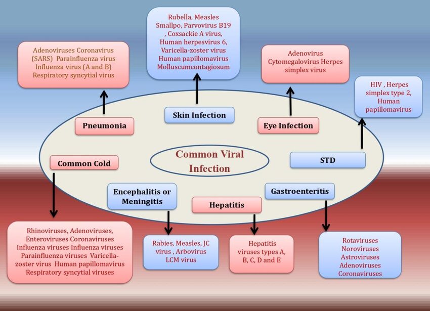
Hemoglobin synthesis depends on adequate iron availability, globin-chain production, and normal heme synthesis. Disorders affecting these processes can produce small red blood cells.
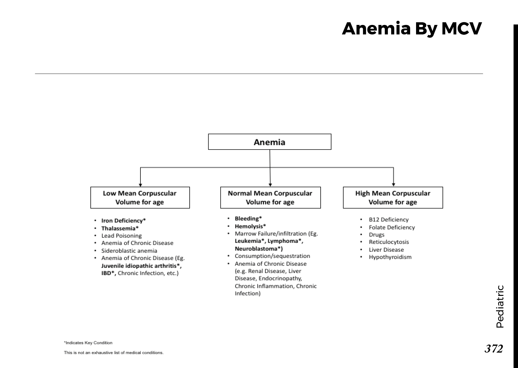
The major diagnostic categories include:
- Iron deficiency anemia.
- Thalassemia and other inherited hemoglobin disorders.
- Anemia associated with chronic inflammation.
- Sideroblastic anemia.
- Lead toxicity in relevant exposure settings.
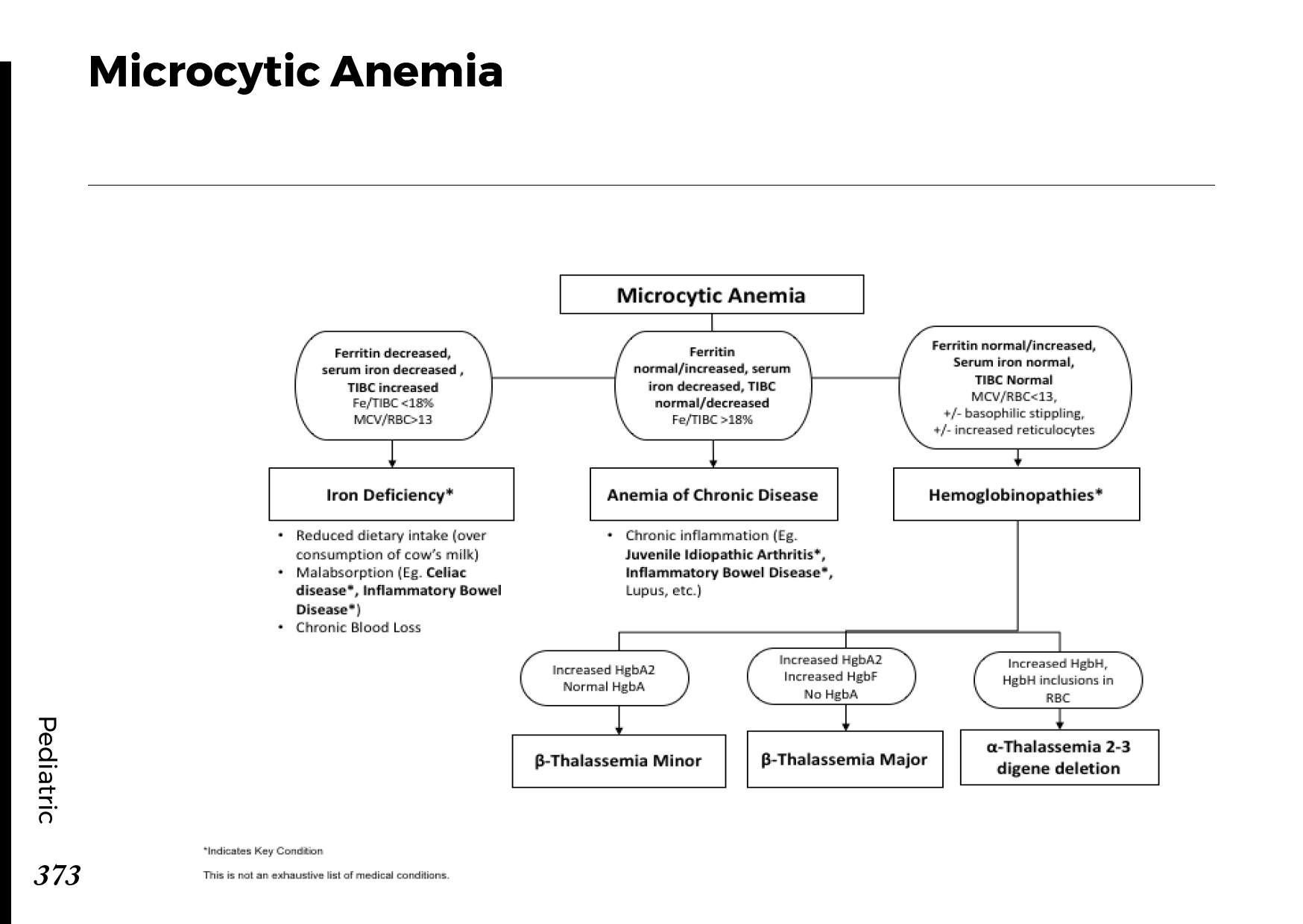
Iron deficiency and thalassemia trait are particularly important to distinguish because both may produce marked microcytosis, but their underlying mechanisms and management differ.
2. Iron Deficiency Anemia: Why Red Cells Become Small
Iron is required for heme synthesis, which is essential for hemoglobin production. When iron stores become depleted, the bone marrow cannot produce red blood cells with their normal hemoglobin content.
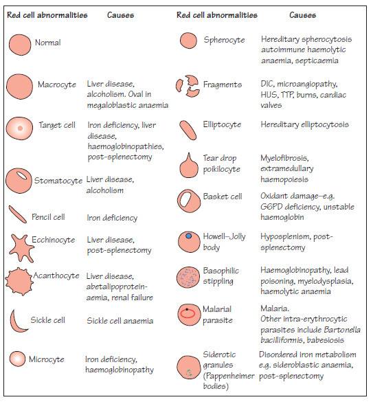
As the deficiency progresses, red cells often become smaller and paler, producing microcytosis and hypochromia.
Common causes
- Chronic blood loss, including heavy menstrual bleeding and gastrointestinal bleeding.
- Inadequate dietary iron intake.
- Increased iron requirements, such as during pregnancy.
- Reduced iron absorption, including selected gastrointestinal disorders and previous gastrointestinal surgery.
- Combined nutritional deficiencies or other conditions that complicate the blood count.
The cause of iron deficiency is as important as confirming the deficiency itself. In adult men and postmenopausal women, unexplained iron deficiency often warrants investigation for gastrointestinal blood loss. The appropriate evaluation depends on age, symptoms, risk factors, and clinical guidelines.
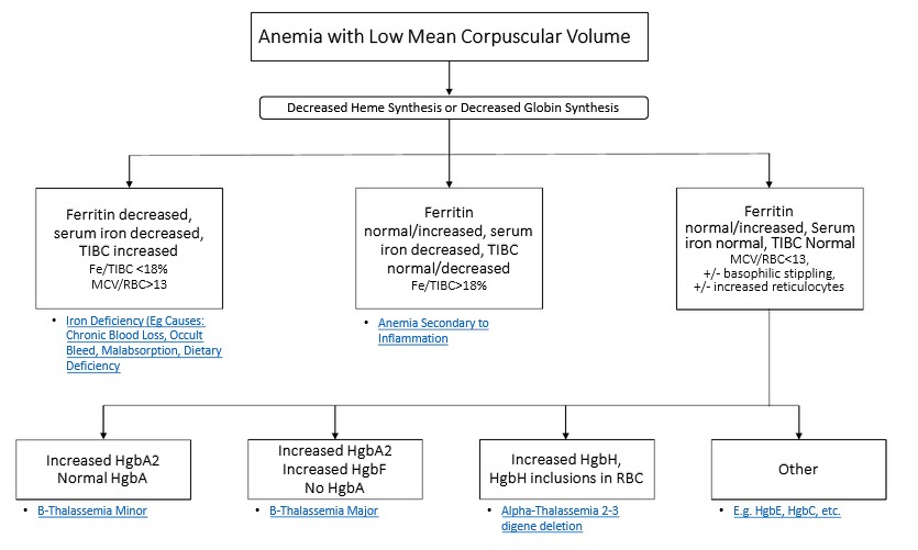
Typical laboratory pattern
- Low hemoglobin.
- Low MCV, particularly in established deficiency.
- Low ferritin in uncomplicated iron deficiency.
- Low serum iron and often increased total iron-binding capacity.
- Increased RDW in many cases as red cell sizes become more variable.
These are typical patterns rather than absolute rules. Early iron deficiency may occur before marked microcytosis develops.
3. Thalassemia Trait: When Microcytosis Is Inherited
Thalassemias are inherited disorders characterized by reduced production of one or more globin chains. Imbalanced globin-chain synthesis affects red cell production and survival.
People with thalassemia trait may have pronounced microcytosis despite having only mild anemia or, in some cases, a hemoglobin concentration within the reference range.
Unlike iron deficiency, thalassemia trait does not necessarily reflect depleted iron stores. Giving iron without evidence of deficiency will not correct the inherited globin-production problem.
Clues on the complete blood count
- MCV that is disproportionately low relative to the degree of anemia.
- A red blood cell count that is relatively preserved or elevated.
- RDW that may be normal or only mildly increased.
- A personal or family history of microcytosis or a known hemoglobin disorder.
These findings raise suspicion but cannot confirm thalassemia. Iron studies and, when indicated, hemoglobin analysis or molecular testing help establish the diagnosis.
4. The Laboratory Clues That Help Separate the Two
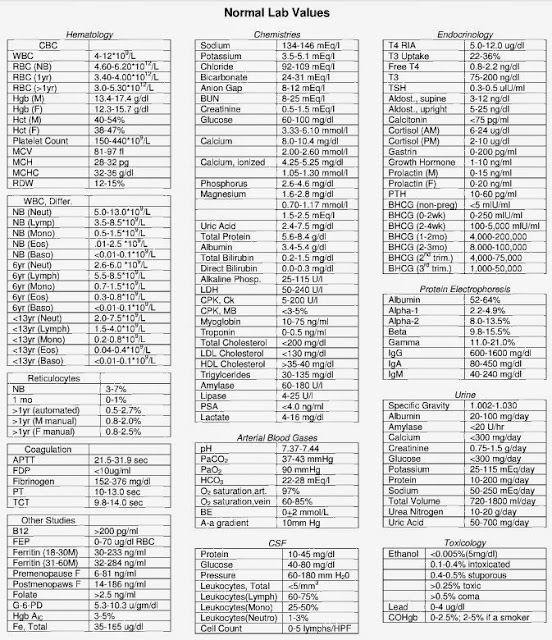
The CBC provides useful clues, but iron studies are generally more informative when determining whether iron deficiency is present.
| Finding | Iron deficiency anemia | Thalassemia trait |
|---|---|---|
| MCV | Low; may decline as deficiency progresses | Often disproportionately low relative to anemia |
| RBC count | Often low or normal | Often relatively preserved or elevated |
| RDW | Often increased | May be normal or mildly increased |
| Ferritin | Usually low in uncomplicated iron deficiency | Usually not low unless iron deficiency coexists |
| Hemoglobin analysis | Does not establish iron deficiency | May identify beta-thalassemia trait or another hemoglobin variant |
5. Ferritin: A Valuable Test With Important Limitations
Ferritin reflects iron stores and is usually the most useful initial laboratory marker for iron deficiency. A clearly low ferritin concentration strongly supports depleted iron stores.
However, ferritin is also an acute-phase reactant. Inflammation, infection, liver disease, and some chronic illnesses can raise ferritin independently of the body's available iron stores.
Therefore, a normal ferritin result does not always exclude iron deficiency in a patient with inflammation or chronic disease.
Depending on the clinical context, clinicians may also review transferrin saturation, serum iron, total iron-binding capacity, inflammatory markers, renal function, and the patient's history.
Why serum iron alone can mislead
Serum iron varies with biological factors and can be reduced in both iron deficiency and inflammation-associated anemia. It should not be interpreted in isolation.
The overall pattern of iron studies, rather than a single measurement, provides a more reliable basis for interpretation.
6. Other Causes of Microcytosis Worth Remembering
Anemia of chronic inflammation
Chronic inflammatory conditions can alter iron regulation through hepcidin and related pathways, restricting iron availability for red blood cell production. The MCV may be normal or low.
Ferritin may be normal or elevated, while serum iron and transferrin saturation may be reduced.
Sideroblastic anemia
Sideroblastic anemia involves impaired heme synthesis and abnormal iron accumulation in erythroid precursors. Causes include inherited disorders, certain medications or toxins, and myelodysplastic conditions. The MCV can vary.
Lead toxicity
Lead exposure can impair heme synthesis and cause anemia. Consider exposure history and appropriate testing when the clinical context suggests this possibility.
Persistent unexplained microcytosis, unusual blood film findings, or discordant iron studies may justify additional hematological evaluation.
7. Clinical Cases: Apply the Reasoning
Case 1: Fatigue and heavy menstrual bleeding
A 29-year-old patient reports fatigue and heavy menstrual periods. Hemoglobin is 9.8 g/dL, MCV is 70 fL, and ferritin is clearly below the laboratory reference range.
Most likely explanation: Iron deficiency anemia.
Next step: Evaluate the cause of iron loss, assess the patient's overall condition, and choose appropriate iron replacement and follow-up. Persistent or disproportionate blood loss requires further investigation.
Case 2: Marked microcytosis with a preserved RBC count
A 22-year-old patient has an MCV of 65 fL, mild anemia, a relatively high red blood cell count, and ferritin within the expected range.
Possible explanation: Thalassemia trait is an important consideration.
Next step: Review the family history and blood count, consider hemoglobin analysis, and investigate further when appropriate. A normal ferritin alone does not prove thalassemia.
Case 3: Microcytosis in a patient with chronic inflammation
A patient with a chronic inflammatory disorder has low serum iron, reduced transferrin saturation, and a ferritin concentration that is not low.
Possible explanation: Inflammation-associated iron restriction is possible, although concurrent iron deficiency cannot be excluded automatically.
Next step: Interpret the iron studies alongside inflammatory activity, clinical history, and any additional tests needed to distinguish absolute iron deficiency from functional iron restriction.
8. A Practical Diagnostic Approach
- Confirm the anemia and microcytosis. Review hemoglobin, MCV, previous CBC results, and the appropriate reference ranges.
- Review the RBC count and RDW. Use these as clues, not definitive diagnostic criteria.
- Order iron studies. Ferritin is a useful starting point; interpret it with inflammatory status and other iron indices when needed.
- Investigate the cause of iron deficiency. Consider blood loss, dietary intake, malabsorption, pregnancy, and other relevant factors.
- Consider hemoglobin analysis. If thalassemia or another hemoglobin disorder is suspected, choose appropriate testing. Iron deficiency can affect interpretation of some hemoglobin fractions.
- Expand the differential when necessary. Persistent unexplained microcytosis may require evaluation for inflammation-associated anemia, lead exposure, sideroblastic anemia, or other disorders.
The aim is not simply to label the red cells as small. It is to identify the mechanism, find any treatable underlying cause, and avoid unnecessary or inappropriate treatment.
Frequently Asked Questions
Can thalassemia trait cause severe microcytosis with mild anemia?
Yes. Thalassemia trait can produce a disproportionately low MCV even when the hemoglobin reduction is relatively modest. Iron studies and appropriate hemoglobin testing help clarify the cause.
Does a normal ferritin exclude iron deficiency?
Not in every situation. Inflammation, infection, and liver disease can increase ferritin, potentially masking depleted or unavailable iron. Additional iron indices may be needed.
Should everyone with microcytosis take iron?
No. Iron treatment is appropriate when iron deficiency is established or sufficiently supported by clinical assessment. Unnecessary supplementation does not correct thalassemia trait and can delay the correct diagnosis.
Can iron deficiency and thalassemia occur together?
Yes. A patient with thalassemia trait can also develop iron deficiency. Finding one condition should not prevent investigation for the other when the results or clinical history suggest both.
When should unexplained microcytosis be investigated further?
Further assessment is appropriate when iron studies do not explain the findings, microcytosis persists, there are concerning symptoms or blood film abnormalities, or an inherited blood disorder is suspected.
Key Takeaways
- Microcytosis is a laboratory finding, not a final diagnosis.
- Iron deficiency and thalassemia trait are common causes but require different approaches.
- RBC count and RDW provide useful clues, but neither can establish the diagnosis alone.
- Ferritin is valuable, although inflammation can complicate its interpretation.
- Investigating the underlying cause is essential, especially when iron deficiency may reflect ongoing blood loss.
- Persistent unexplained microcytosis may warrant hemoglobin analysis or further hematological assessment.
 Menstrual cycle physiology: