Pulmonary Embolism: Recognizing the Clues That Are Easy to Miss
A practical clinical guide for medical students and healthcare professionals.
A patient presents with sudden shortness of breath, a rapid heart rate, and vague chest discomfort. The oxygen saturation may be only mildly reduced, the lungs may sound clear, and the initial chest radiograph may show no obvious abnormality. Could this still be a life-threatening emergency?
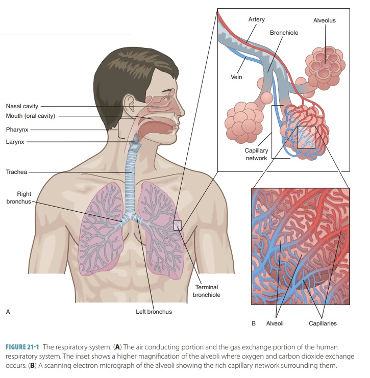
Pulmonary embolism (PE) occurs when material, usually a blood clot, obstructs the pulmonary arterial circulation. Because its symptoms overlap with many common conditions, the diagnosis depends on recognizing risk factors, estimating clinical probability, and selecting investigations appropriately.
1. Why Pulmonary Embolism Can Be Difficult to Recognize
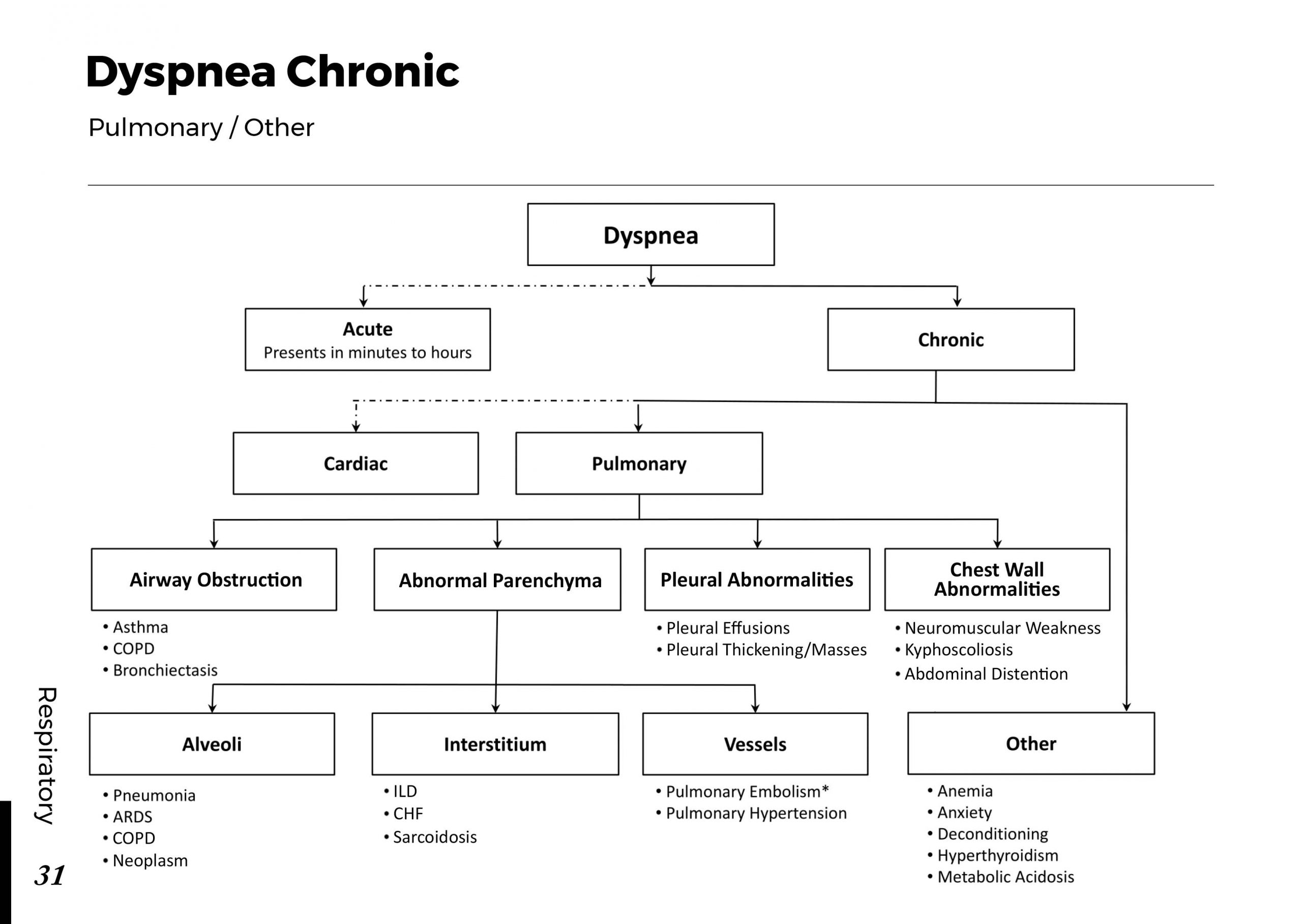
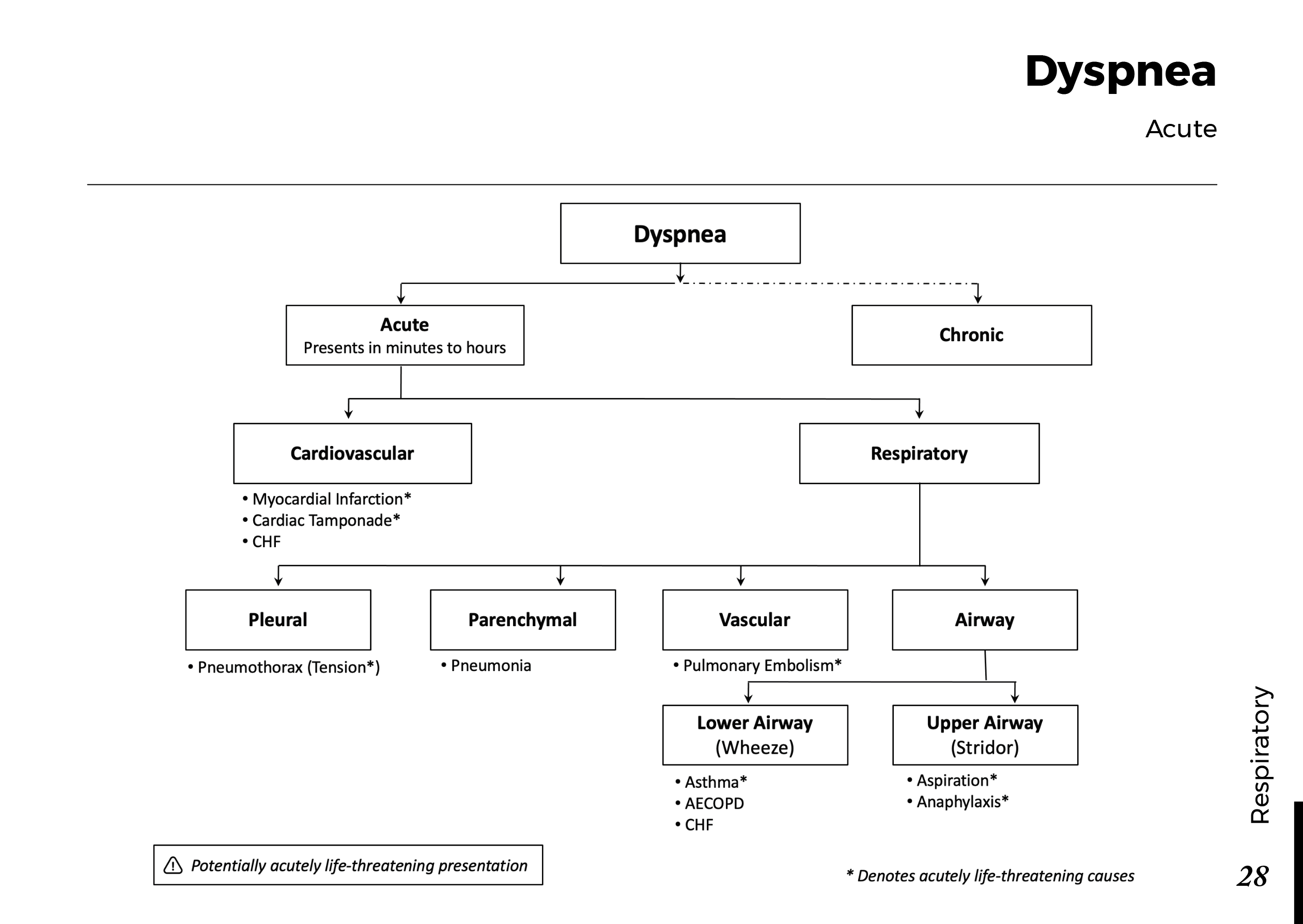
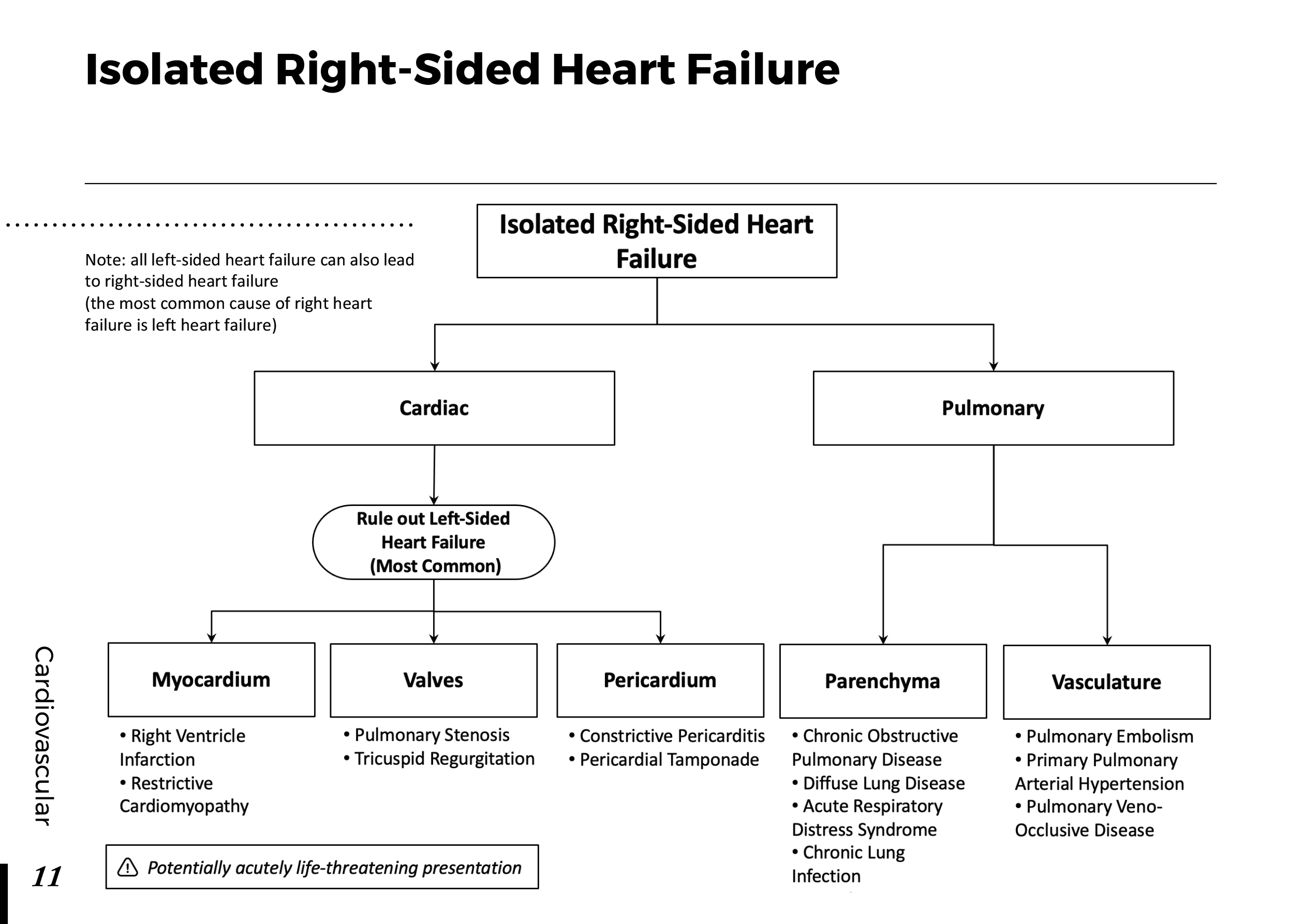
PE does not have one characteristic presentation. Some patients develop sudden, severe respiratory symptoms, while others report gradually worsening breathlessness, unexplained tachycardia, or discomfort that is initially attributed to another condition.
Common presentations include:
- Sudden or unexplained shortness of breath.
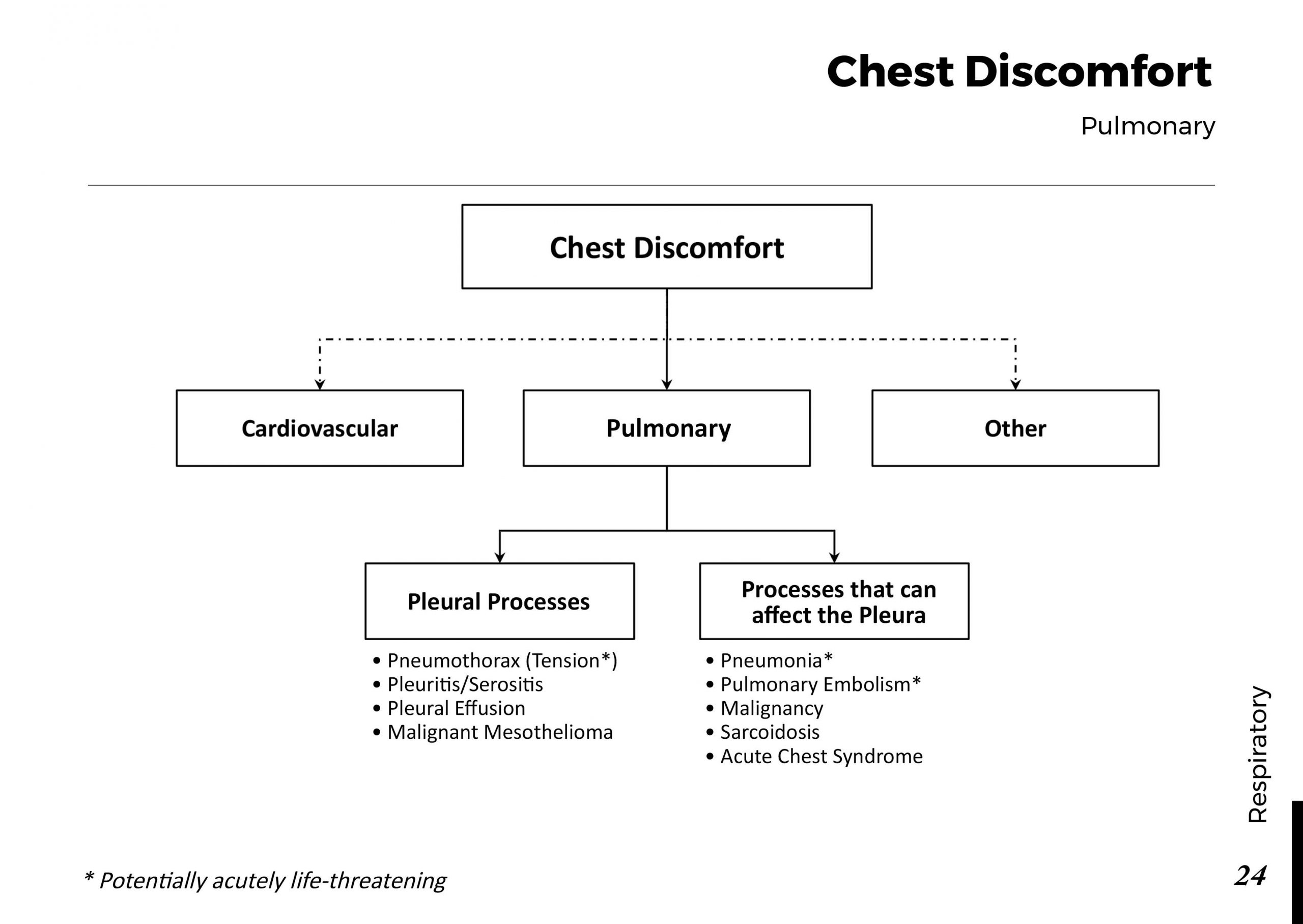
- Pleuritic chest pain that becomes worse with inspiration.
- Tachycardia or an increased respiratory rate.
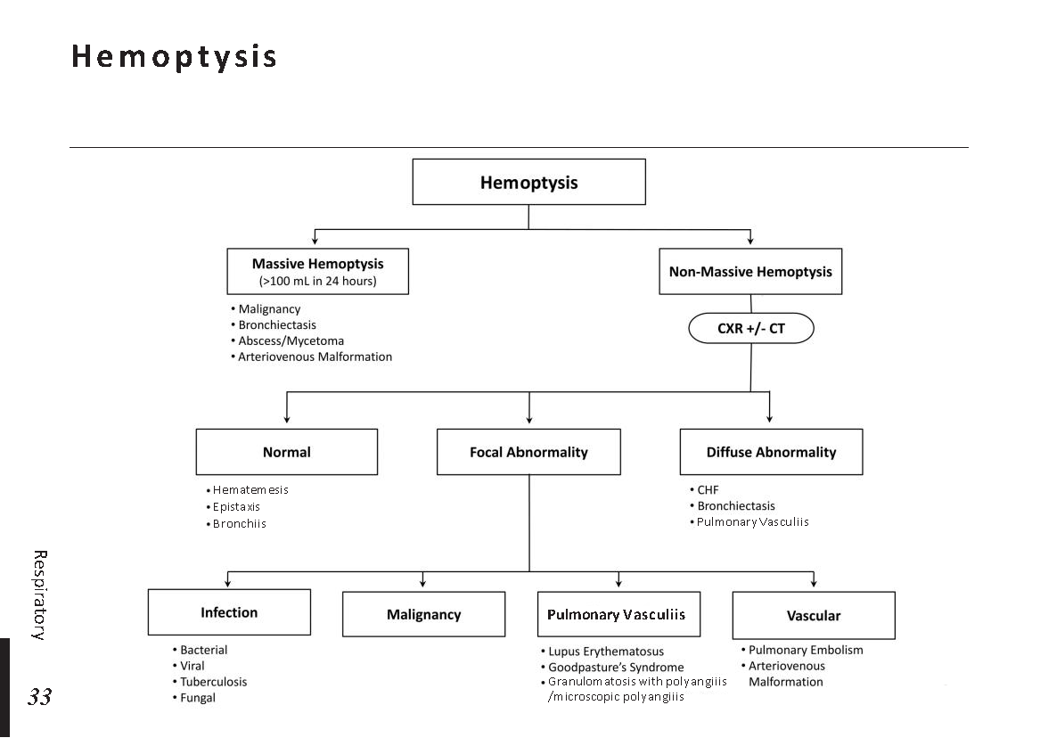
- Cough, occasionally accompanied by hemoptysis.
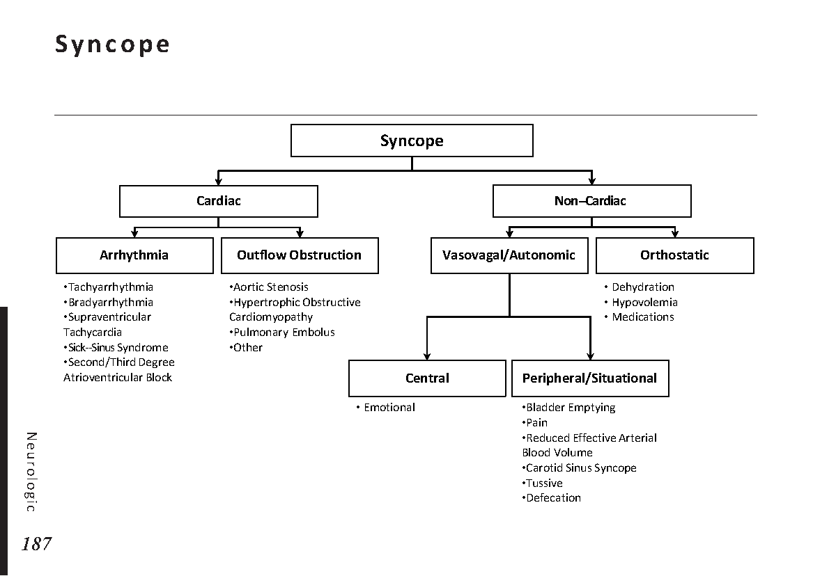
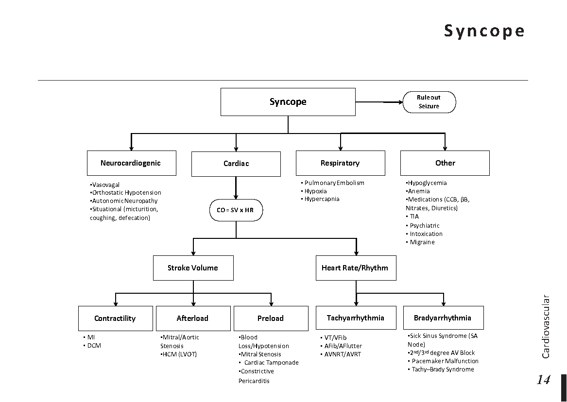
- Syncope or presyncope, particularly in more serious presentations.
- Unilateral leg swelling or pain suggesting deep vein thrombosis (DVT).
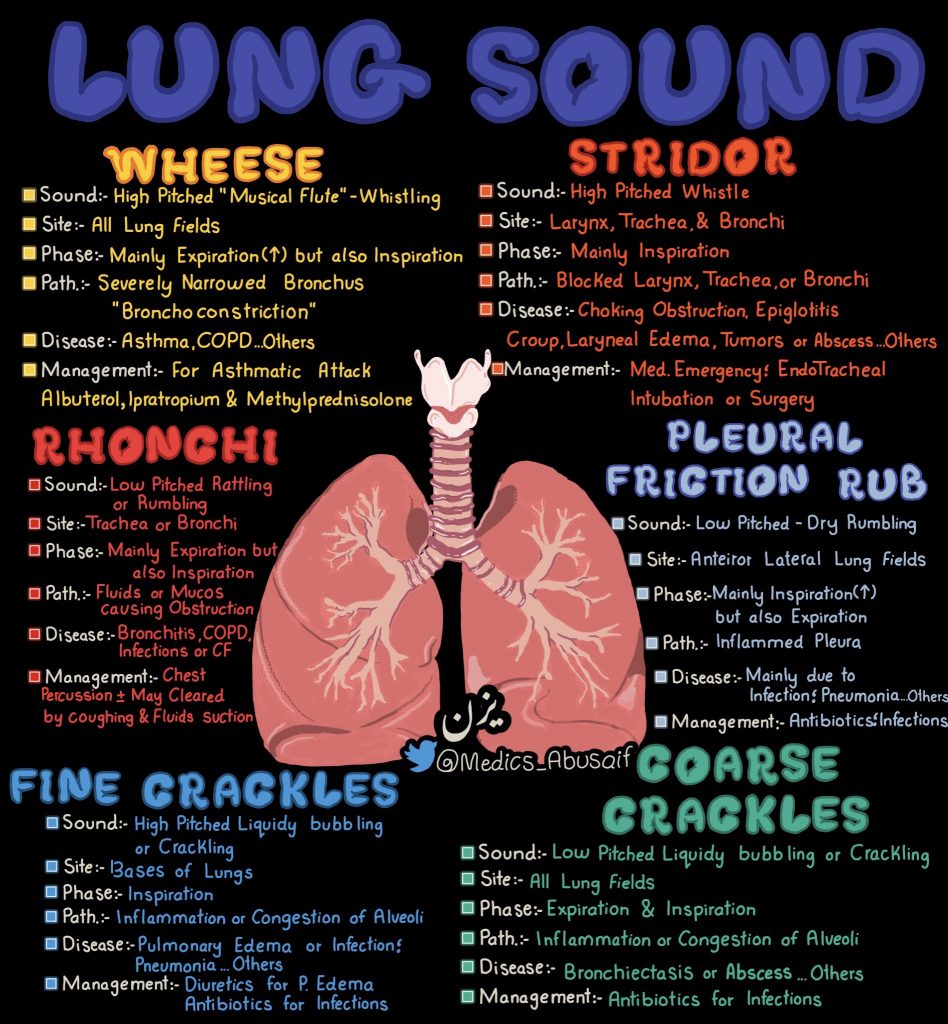
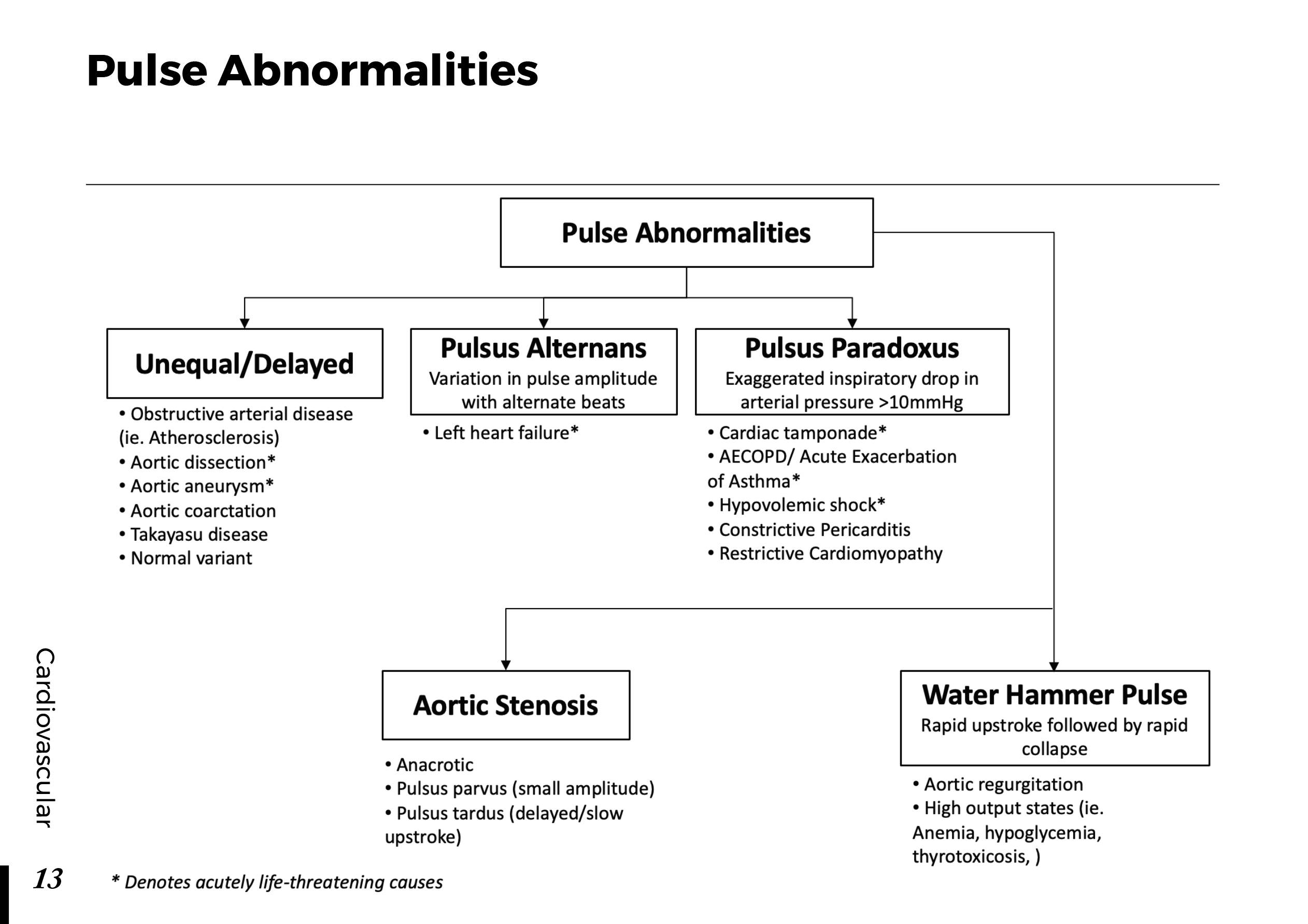
Examination may reveal tachypnea, tachycardia, hypoxemia, signs of DVT, or evidence of circulatory compromise. However, normal lung auscultation and a relatively unremarkable chest radiograph do not exclude PE.
The absence of dramatic symptoms should not create false reassurance when the patient's risk factors and clinical presentation remain concerning.
2. Who Is at Increased Risk?
Most pulmonary emboli originate from thrombi in the deep veins of the lower extremities or pelvis. Virchow's triad provides a useful framework for understanding why venous thrombosis develops.
Venous stasis
Prolonged immobility, hospitalization, major surgery, and extended periods of reduced mobility can impair venous blood flow and increase thrombotic risk.
Endothelial injury
Surgery, trauma, and certain vascular injuries can damage the vessel lining and promote clot formation.
Hypercoagulability
Active cancer, pregnancy and the postpartum period, estrogen-containing medications, and inherited or acquired thrombophilias can increase the tendency to form clots.
Previous DVT or PE is another particularly important risk factor. Nevertheless, PE can occur in patients without an obvious provoking factor, so the absence of a recognized risk factor does not completely exclude the diagnosis.
3. The First Decision: Is the Patient Hemodynamically Unstable?
Before selecting a diagnostic pathway, clinicians must assess the patient's immediate stability.
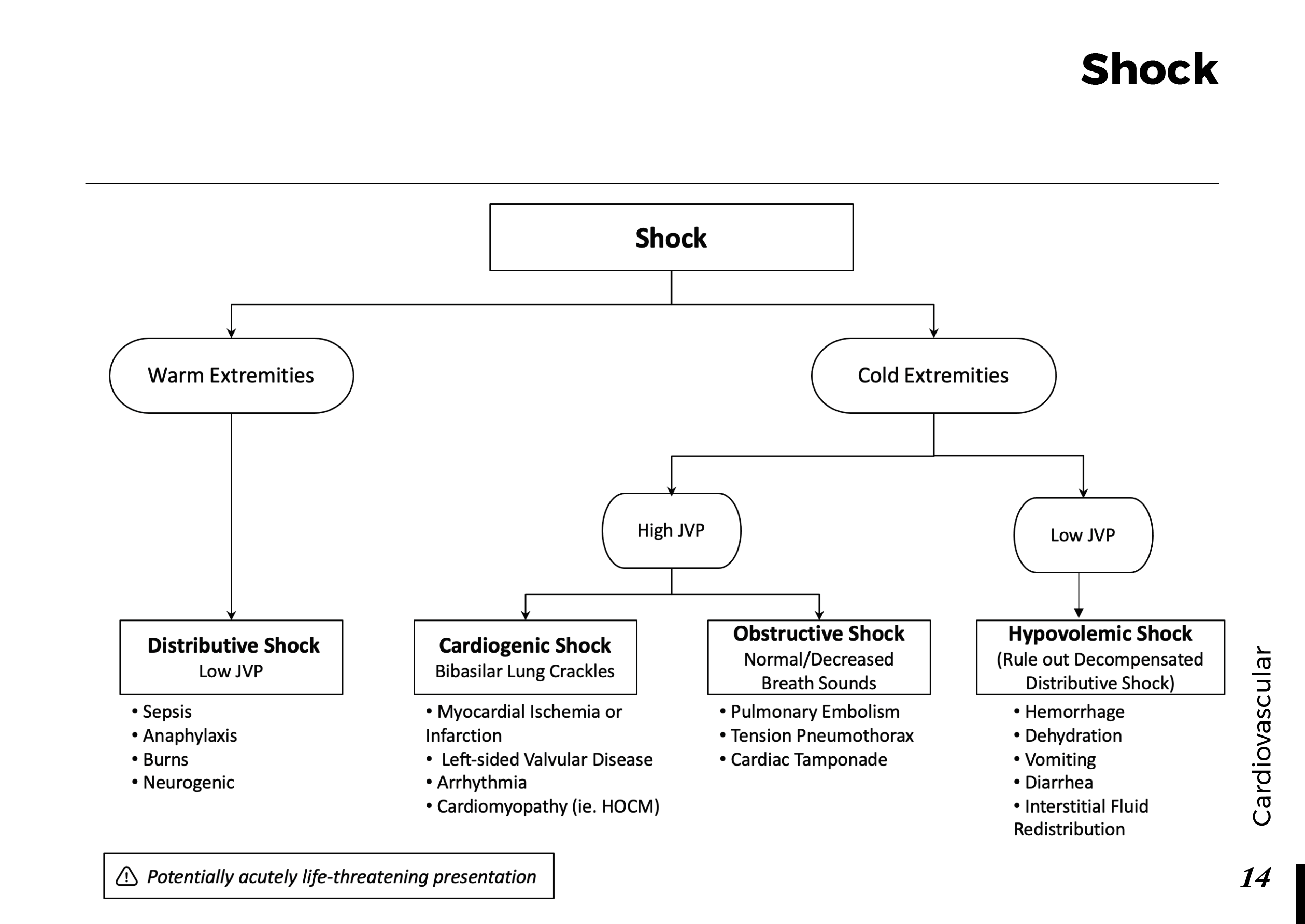
Persistent hypotension, obstructive shock, cardiac arrest, or severe circulatory compromise in a patient with suspected PE raises concern for a high-risk presentation. Such patients require immediate resuscitation and urgent evaluation.
In a sufficiently stable patient, clinicians can generally proceed through a structured assessment of pretest probability followed by appropriate laboratory testing or imaging.
The distinction matters because a diagnostic strategy designed for a stable outpatient should not delay urgent treatment decisions in a patient who is deteriorating.
4. Clinical Probability: Why Wells Score Matters
The Wells score is one tool used to estimate the pretest probability of PE. It combines clinical features and risk factors to help determine which investigation is appropriate.
Its components include:
- Clinical signs of DVT.
- Whether PE is considered more likely than an alternative diagnosis.
- Heart rate above 100 beats per minute.
- Recent surgery or immobilization.
- Previous DVT or PE.
- Hemoptysis.
- Active malignancy.
Different validated versions use different scoring approaches and probability categories. Clinicians should use the version adopted by their institution rather than mixing criteria from different pathways.
The score is not a substitute for judgment. Its purpose is to structure clinical reasoning and help select the next step safely.
5. D-Dimer: Useful When Used in the Right Patient
D-dimer is a degradation product formed when cross-linked fibrin is broken down. It can be elevated in venous thromboembolism, but it is also increased in many other conditions.
Examples include infection, malignancy, pregnancy, recent surgery, trauma, and increasing age. Therefore, a positive result does not establish the diagnosis of PE.
When is D-dimer most helpful?
In patients with an appropriately low or intermediate pretest probability, a negative high-sensitivity D-dimer can help exclude PE when used within a validated diagnostic pathway.
In selected patients aged 50 years or older, an age-adjusted threshold may improve specificity. The exact threshold depends on the assay and reporting units; clinicians must follow the locally validated protocol.
Patients with a high clinical probability generally proceed to appropriate imaging rather than relying on a D-dimer result to exclude PE.
6. Imaging: Confirming or Excluding the Diagnosis
CT pulmonary angiography
CT pulmonary angiography (CTPA) is commonly the preferred imaging test for patients who require diagnostic imaging and can undergo the examination. It can demonstrate filling defects in the pulmonary arteries and may identify alternative explanations for symptoms.
The decision to use CTPA should account for contrast-related concerns, kidney function when relevant, pregnancy, radiation exposure, and the patient's overall condition.
Ventilation-perfusion scanning
A ventilation-perfusion (V/Q) scan is an alternative in selected circumstances, including some patients in whom iodinated contrast is unsuitable. Its interpretation depends partly on the scan pattern and clinical context.
Lower-extremity venous ultrasound
Compression ultrasonography can identify DVT in the legs. Finding a proximal DVT in an appropriate patient may establish venous thromboembolism and influence management, particularly when chest imaging is difficult or unsuitable.
No single imaging approach is appropriate for every patient. The best choice depends on clinical probability, availability, contraindications, and the diagnostic pathway being followed.
7. What Do ECG, Chest X-Ray, and Blood Gases Tell Us?
These tests can provide useful supporting information, but none should be used alone to rule out PE.
Electrocardiogram
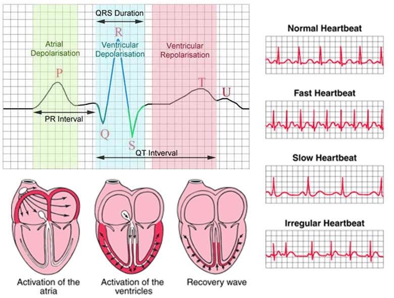
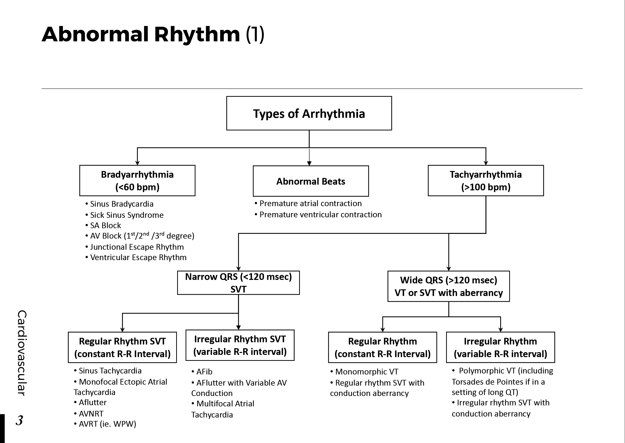
Sinus tachycardia is a common finding. Other abnormalities may occur, including right-heart strain patterns, right bundle branch block, or anterior T-wave inversions. The classic S1Q3T3 pattern is neither sufficiently sensitive nor sufficiently specific to establish the diagnosis.
Chest radiograph
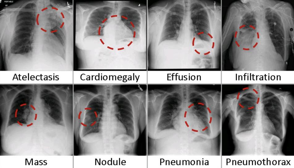
The chest radiograph may be normal or show nonspecific abnormalities. Its role often includes identifying alternative causes of breathlessness and helping determine whether certain imaging approaches are appropriate.
Arterial blood gas
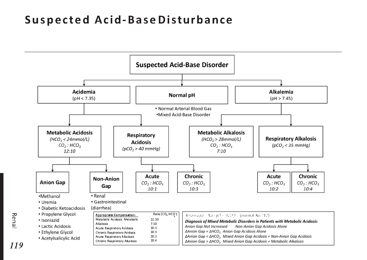
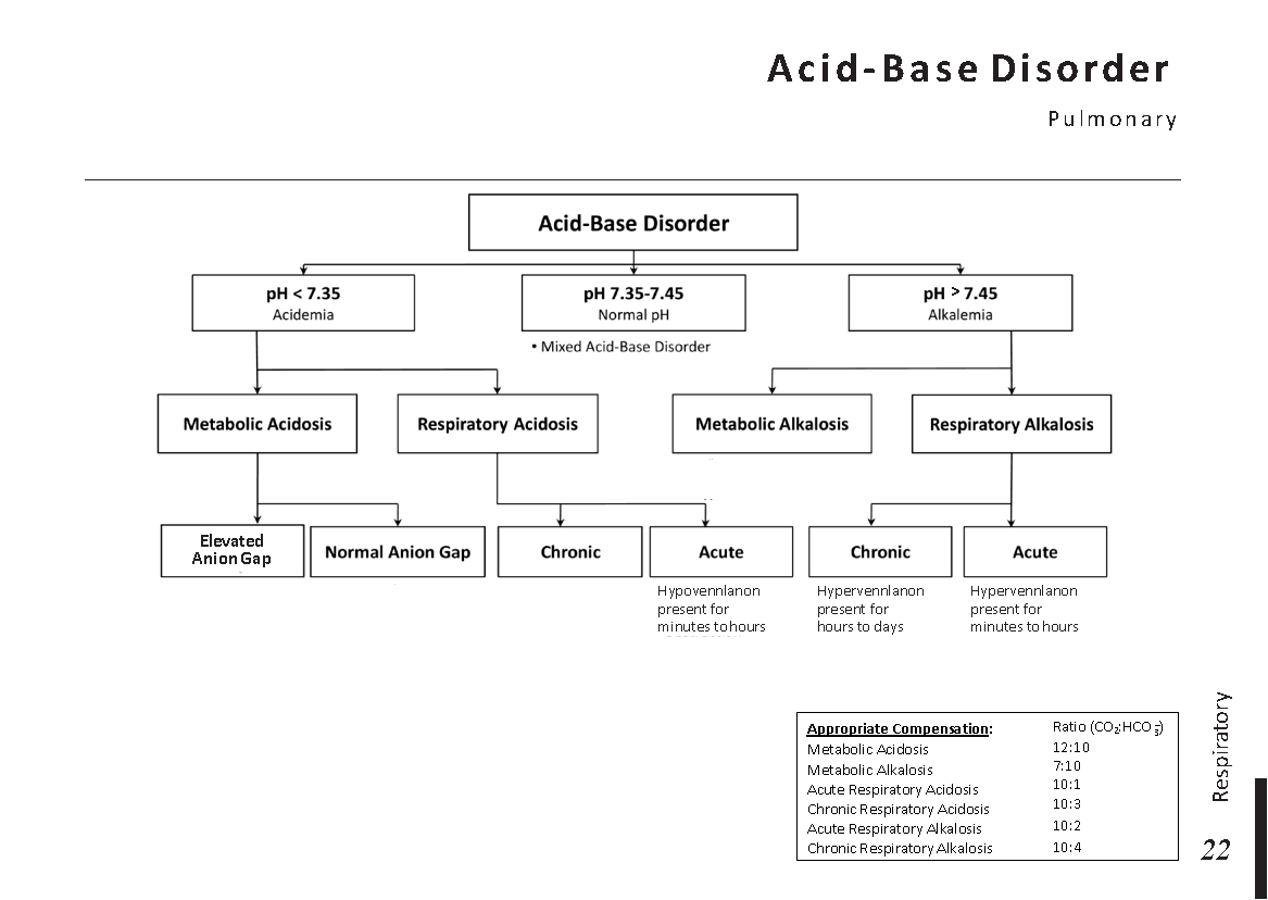
Hypoxemia and respiratory alkalosis may occur because of increased respiratory drive and ventilation-perfusion mismatch. However, blood gas results can also be normal, especially in less severe cases.
These investigations help characterize the presentation and assess severity. They do not replace a probability-based diagnostic strategy.
8. Clinical Cases: Applying the Diagnostic Approach
Case 1: Sudden dyspnea after surgery
A 62-year-old patient develops sudden shortness of breath and tachycardia several days after major orthopedic surgery.
Clinical reasoning: Recent major surgery increases thrombotic risk. PE should be considered urgently, and the patient requires assessment of hemodynamic stability and a suitable diagnostic pathway.
Learning point: A strong provoking risk factor changes clinical probability and may influence the choice of investigation.
Case 2: Pleuritic pain and unilateral leg swelling
A patient presents with pain on inspiration, unexplained breathlessness, and swelling of one lower leg.
Clinical reasoning: The combination raises concern for venous thromboembolism. Leg ultrasound and chest imaging may be selected according to the clinical situation.
Learning point: Findings outside the chest can provide important clues to the cause of respiratory symptoms.
Case 3: Positive D-dimer in a patient with infection
A patient with an acute infection develops mild breathlessness and has an elevated D-dimer.
Clinical reasoning: Infection itself can elevate D-dimer. The result does not confirm PE; the clinician must reassess the pretest probability and decide whether further testing is indicated.
Learning point: Interpreting a test without considering its specificity can lead to unnecessary investigations or incorrect conclusions.
9. Common Diagnostic Mistakes
- Ruling out PE because the chest radiograph is normal. A normal radiograph does not exclude pulmonary vascular obstruction.
- Diagnosing PE from D-dimer alone. An elevated value is nonspecific.
- Using D-dimer indiscriminately. Testing is most useful when selected according to a validated probability-based strategy.
- Over-relying on oxygen saturation. A normal or near-normal reading does not reliably exclude PE.
- Ignoring hemodynamic instability. Shock or persistent hypotension changes the urgency and diagnostic approach.
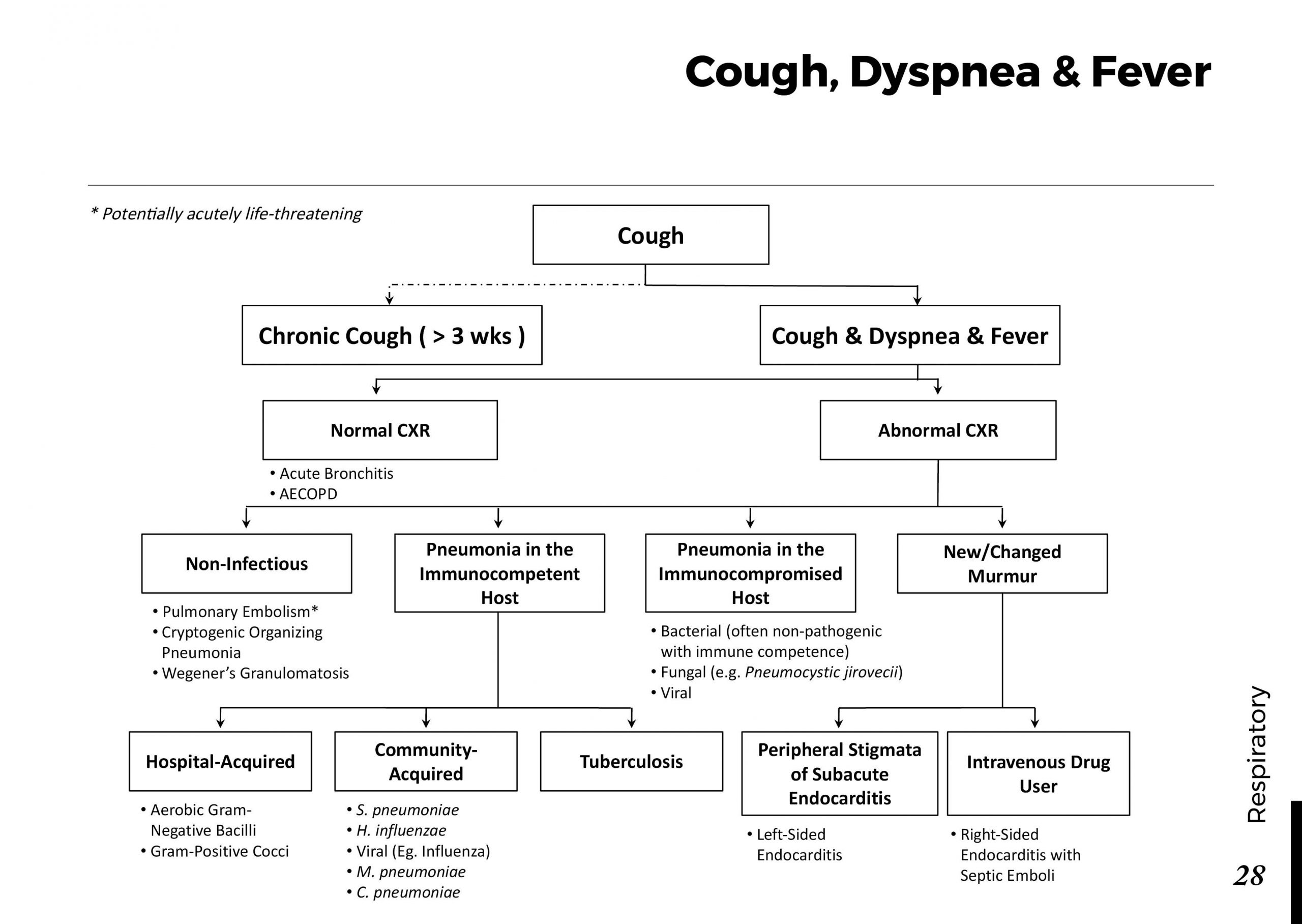
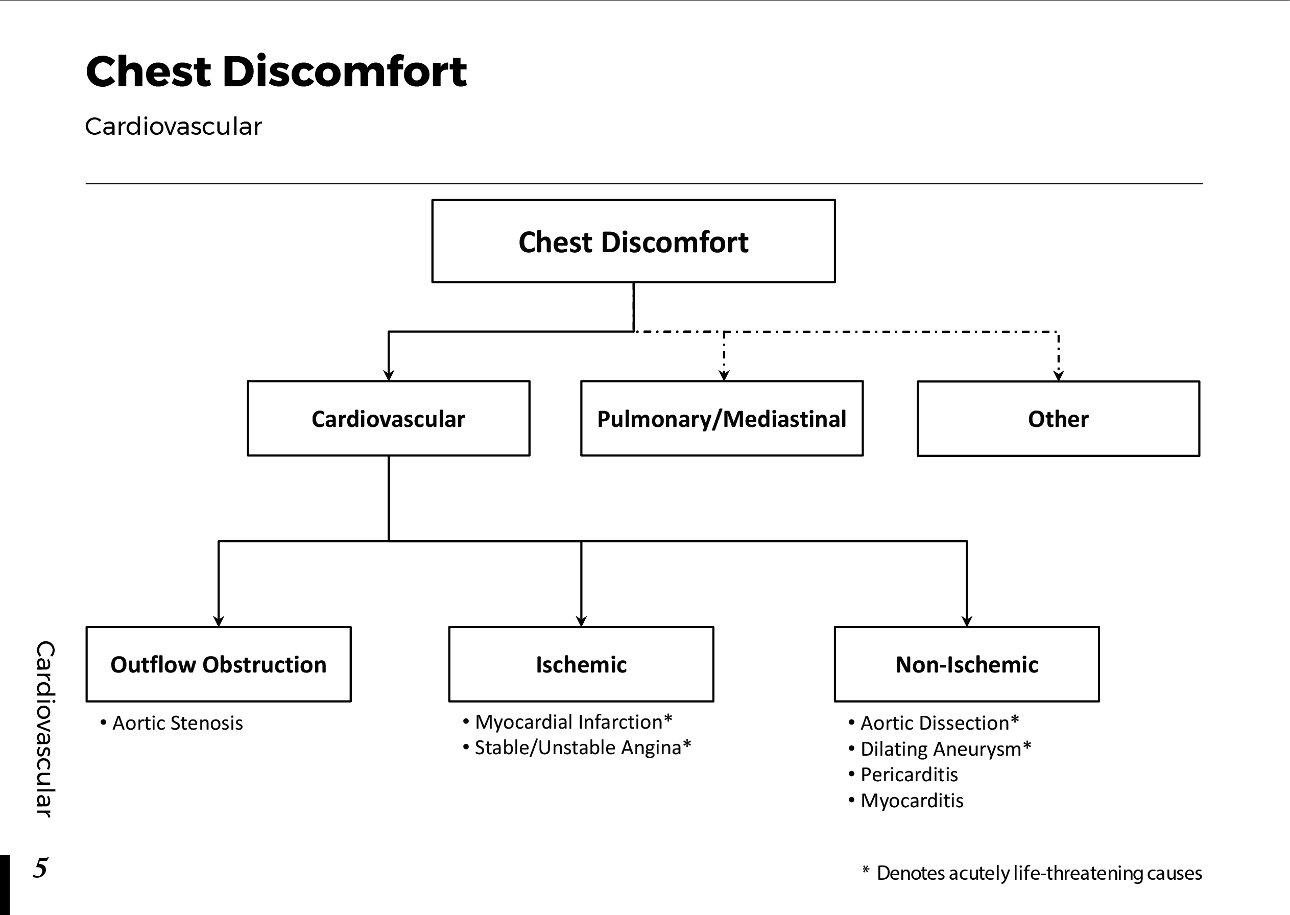
- Forgetting alternative diagnoses. Acute coronary syndrome, pneumonia, pneumothorax, heart failure, and other conditions can produce overlapping symptoms.
Frequently Asked Questions
Can pulmonary embolism occur with a normal oxygen saturation?
Yes. Oxygen saturation can be normal, particularly in less severe cases. It should not be used alone to exclude PE.
Does a normal ECG exclude pulmonary embolism?
No. ECG findings are often nonspecific, and a normal ECG does not reliably rule out PE.
Is a positive D-dimer diagnostic of PE?
No. D-dimer can increase in many conditions. In an appropriate patient, it is primarily used to help exclude PE when the result is negative and the validated diagnostic pathway permits this interpretation.
What is the preferred imaging test for pulmonary embolism?
CTPA is commonly the first-choice imaging examination for suitable patients who require imaging. V/Q scanning and other approaches may be preferable in selected circumstances.
What symptoms require emergency assessment?
Sudden severe breathlessness, chest pain accompanied by collapse, fainting, coughing up blood, or signs of shock require urgent emergency assessment. These symptoms can have several serious causes, including PE.
Key Takeaways
- PE can present with nonspecific symptoms, and the absence of classic findings does not exclude it.
- Prior venous thromboembolism, recent surgery, immobility, cancer, and other risk factors help shape clinical probability.
- Assess hemodynamic stability before choosing the diagnostic pathway.
- Use clinical probability tools and D-dimer selectively according to validated protocols.
- CTPA is commonly used to confirm or exclude PE when imaging is indicated and appropriate.
- ECG, chest radiography, and blood gas results provide supporting information but cannot independently rule out PE.
 Menstrual cycle physiology: